• 文献检索
  • 文档翻译
  • 深度研究
  • 学术资讯
  • Suppr Zotero 插件Zotero 插件
  • 邀请有礼
  • 套餐&价格
  • 历史记录
应用&插件
Suppr Zotero 插件Zotero 插件浏览器插件Mac 客户端Windows 客户端微信小程序
定价
高级版会员购买积分包购买API积分包
服务
文献检索文档翻译深度研究API 文档MCP 服务
关于我们
关于 Suppr公司介绍联系我们用户协议隐私条款
关注我们

Suppr 超能文献

核心技术专利:CN118964589B侵权必究
粤ICP备2023148730 号-1Suppr @ 2026

文献检索

告别复杂PubMed语法,用中文像聊天一样搜索,搜遍4000万医学文献。AI智能推荐,让科研检索更轻松。

立即免费搜索

文件翻译

保留排版,准确专业,支持PDF/Word/PPT等文件格式,支持 12+语言互译。

免费翻译文档

深度研究

AI帮你快速写综述,25分钟生成高质量综述,智能提取关键信息,辅助科研写作。

立即免费体验

单细胞转录组学为成纤维细胞在腹膜纤维化过程中的作用提供了新的见解。

Single-cell transcriptomics provides new insights into the role of fibroblasts during peritoneal fibrosis.

作者信息

Zhang Jinhua, Chen Yuxian, Chen Tufeng, Miao Bin, Tang Zuofu, Hu Xiao, Luo You, Zheng Tong, Na Ning

机构信息

Department of Kidney Transplantation, The Third Affiliated Hospital of Sun Yat-sen University, Guangzhou, China.

Department of Joint Surgery, The Third Affiliated Hospital of Sun Yat-sen University, Guangzhou, China.

出版信息

Clin Transl Med. 2021 Mar;11(3):e321. doi: 10.1002/ctm2.321.

DOI:10.1002/ctm2.321
PMID:33784014
原文链接:https://pmc.ncbi.nlm.nih.gov/articles/PMC7908046/
Abstract

BACKGROUND

The contributions of various types of cell populations in dialysis-related peritoneal fibrosis are poorly understood. Single-cell RNA sequencing brings single-cell level resolution to the analysis of cellular transcriptomics, which provides a new way to further characterize the distinct roles and functional states of each cell population during peritoneal fibrosis.

METHODS

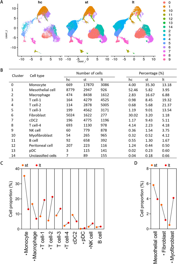
Single-cell transcriptomics from normal peritoneal tissues of six patients, from effluent of patients with short-term peritoneal dialysis (less than 2 weeks, n = 6), and from long-term peritoneal dialysis patients (more than 6 years, n = 4) were analyzed.

RESULTS

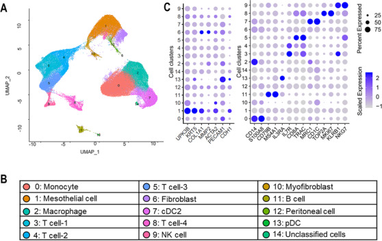
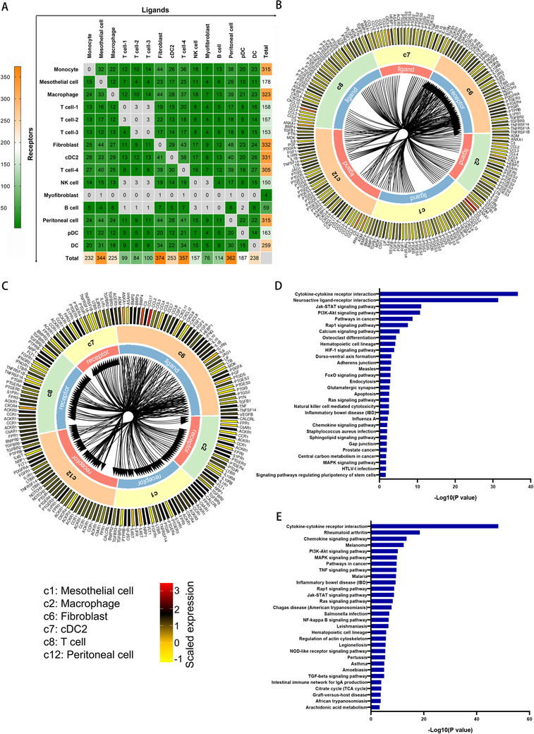
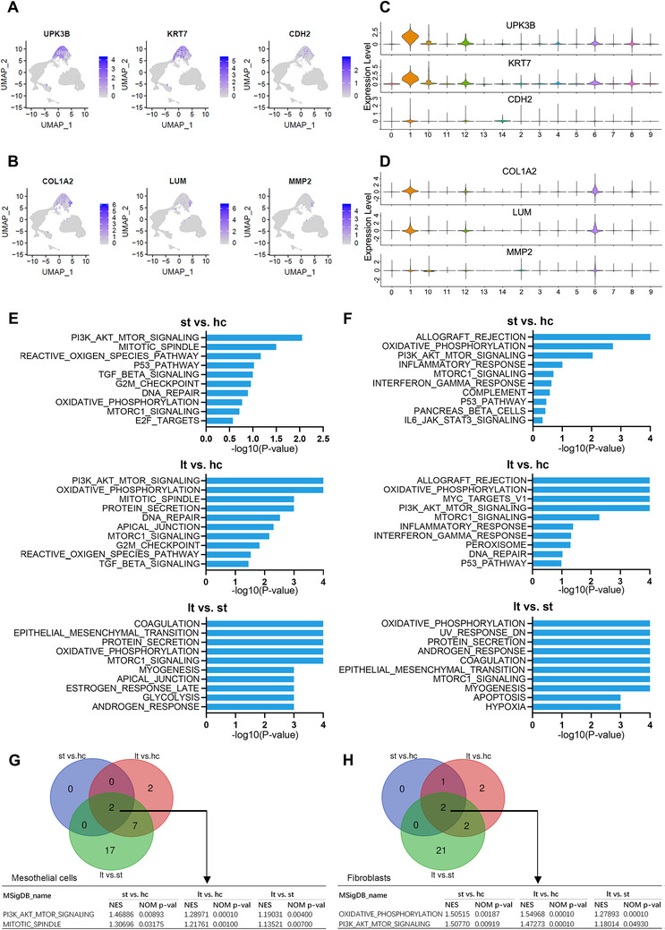
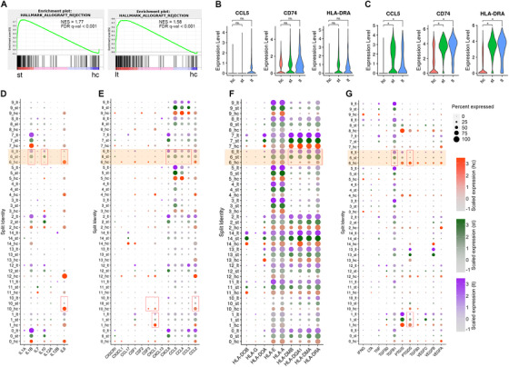
We identified a distinct cell component between samples among different groups. Functional analysis of the differentially expressed genes identified cell type specific biological processes relevant to different fibrosis stages. Well-known key molecular mechanisms participating in the pathophysiology of peritoneal fibrosis were vitrified, and some of them were found to be restricted to specific cell types. Gradually growing enrichment of PI3K/AKT/mTOR pathway and impairment of oxidative phosphorylation in mesothelial cells and fibroblasts were found from healthy control, short-term dialysis, to long-term dialysis, respectively. The fibroblasts' population obtained from the patients, who received peritoneal dialysis, showed a functional characteristic of immune-chemotaxis and immune response, which was characterized by broadly significant increase in the expression of interleukins, chemokines, cytokines, and human leukocyte antigens. Furthermore, we described the intercellular crosstalk networks based on receptor-ligand interactions, and highlighted a central role of fibroblasts in regulating the key mechanisms of peritoneal fibrosis through crosstalk with other cells.

CONCLUSIONS

In summary, despite describing information for fibrogenic molecular mechanisms in the resolution level of individual cell populations, this work identifies the significant functional evolution of fibroblasts during peritoneal fibrosis. This study also reveals the intercellular receptor-ligand interactions in which the fibroblasts serve as a major node, eventually providing new insights into the role of fibroblasts during disease pathogenesis.

摘要

背景

目前对透析相关腹膜纤维化中各类细胞群的作用了解甚少。单细胞RNA测序为细胞转录组学分析带来了单细胞水平的分辨率,为进一步明确腹膜纤维化过程中每个细胞群的独特作用和功能状态提供了新途径。

方法

对6例患者正常腹膜组织、短期腹膜透析患者(少于2周,n = 6)腹透液以及长期腹膜透析患者(超过6年,n = 4)的单细胞转录组进行分析。

结果

我们在不同组的样本间鉴定出了不同的细胞成分。对差异表达基因的功能分析确定了与不同纤维化阶段相关的细胞类型特异性生物学过程。参与腹膜纤维化病理生理学的一些知名关键分子机制得到了验证,其中一些机制被发现局限于特定细胞类型。从健康对照到短期透析再到长期透析,分别发现间皮细胞和成纤维细胞中PI3K/AKT/mTOR通路逐渐富集以及氧化磷酸化受损。接受腹膜透析患者的成纤维细胞群表现出免疫趋化和免疫反应的功能特征,其特征是白细胞介素、趋化因子、细胞因子和人类白细胞抗原的表达普遍显著增加。此外,我们基于受体-配体相互作用描述了细胞间串扰网络,并强调了成纤维细胞在通过与其他细胞串扰来调节腹膜纤维化关键机制中的核心作用。

结论

总之,尽管本研究在单个细胞群水平描述了纤维化分子机制的信息,但确定了腹膜纤维化过程中成纤维细胞的显著功能演变。本研究还揭示了以成纤维细胞为主要节点的细胞间受体-配体相互作用,最终为成纤维细胞在疾病发病机制中的作用提供了新见解。

https://cdn.ncbi.nlm.nih.gov/pmc/blobs/76c7/7908046/69f1f89abba7/CTM2-11-e321-g005.jpg
https://cdn.ncbi.nlm.nih.gov/pmc/blobs/76c7/7908046/52d3dcd2cd8b/CTM2-11-e321-g001.jpg
https://cdn.ncbi.nlm.nih.gov/pmc/blobs/76c7/7908046/57428ef6b61d/CTM2-11-e321-g002.jpg
https://cdn.ncbi.nlm.nih.gov/pmc/blobs/76c7/7908046/117b1a5a38a2/CTM2-11-e321-g003.jpg
https://cdn.ncbi.nlm.nih.gov/pmc/blobs/76c7/7908046/2a40a954b996/CTM2-11-e321-g004.jpg
https://cdn.ncbi.nlm.nih.gov/pmc/blobs/76c7/7908046/69f1f89abba7/CTM2-11-e321-g005.jpg

相似文献

1
Single-cell transcriptomics provides new insights into the role of fibroblasts during peritoneal fibrosis.单细胞转录组学为成纤维细胞在腹膜纤维化过程中的作用提供了新的见解。
Clin Transl Med. 2021 Mar;11(3):e321. doi: 10.1002/ctm2.321.
2
TGF-β1-VEGF-A pathway induces neoangiogenesis with peritoneal fibrosis in patients undergoing peritoneal dialysis.TGF-β1-VEGF-A 通路诱导接受腹膜透析患者的新生血管形成和腹膜纤维化。
Am J Physiol Renal Physiol. 2018 Feb 1;314(2):F167-F180. doi: 10.1152/ajprenal.00052.2017. Epub 2017 Oct 4.
3
Transcriptional patterns in peritoneal tissue of encapsulating peritoneal sclerosis, a complication of chronic peritoneal dialysis.慢性腹膜透析并发症包裹性腹膜硬化症腹膜组织中的转录模式。
PLoS One. 2013;8(2):e56389. doi: 10.1371/journal.pone.0056389. Epub 2013 Feb 13.
4
Preventing peritoneal membrane fibrosis in peritoneal dialysis patients.预防腹膜透析患者腹膜纤维化。
Kidney Int. 2016 Sep;90(3):515-24. doi: 10.1016/j.kint.2016.03.040. Epub 2016 Jun 7.
5
Pleiotrophin triggers inflammation and increased peritoneal permeability leading to peritoneal fibrosis.pleiotrophin 触发炎症和增加腹膜通透性,导致腹膜纤维化。
Kidney Int. 2012 Jan;81(2):160-9. doi: 10.1038/ki.2011.305. Epub 2011 Aug 31.
6
Extracellular vesicle-packaged ILK from mesothelial cells promotes fibroblast activation in peritoneal fibrosis.细胞外小泡包裹的间质细胞中的整合素连接激酶促进腹膜纤维化中的成纤维细胞活化。
J Extracell Vesicles. 2023 Jul;12(7):e12334. doi: 10.1002/jev2.12334.
7
Periostin: a matricellular protein involved in peritoneal injury during peritoneal dialysis.骨膜蛋白:一种细胞外基质蛋白,参与腹膜透析过程中的腹膜损伤。
Perit Dial Int. 2013 Sep-Oct;33(5):515-28. doi: 10.3747/pdi.2010.00259. Epub 2013 Feb 1.
8
Strategies for preventing peritoneal fibrosis in peritoneal dialysis patients: new insights based on peritoneal inflammation and angiogenesis.预防腹膜透析患者腹膜纤维化的策略:基于腹膜炎症和血管生成的新见解。
Front Med. 2017 Sep;11(3):349-358. doi: 10.1007/s11684-017-0571-2. Epub 2017 Aug 8.
9
Alternative activation of macrophages in human peritoneum: implications for peritoneal fibrosis.人腹膜中巨噬细胞的交替激活:对腹膜纤维化的影响。
Nephrol Dial Transplant. 2011 Sep;26(9):2995-3005. doi: 10.1093/ndt/gfq771. Epub 2011 Feb 15.
10
HIF1A and VEGF regulate each other by competing endogenous RNA mechanism and involve in the pathogenesis of peritoneal fibrosis.HIF1A和VEGF通过竞争性内源RNA机制相互调节,并参与腹膜纤维化的发病机制。
Pathol Res Pract. 2019 Apr;215(4):644-652. doi: 10.1016/j.prp.2018.12.022. Epub 2018 Dec 26.

引用本文的文献

1
Integrating single-cell RNA-seq, bulk RNA-seq and network pharmacology reveals protective effect of salidroside in peritoneal dialysis-associated peritoneal fibrosis.整合单细胞RNA测序、批量RNA测序和网络药理学揭示了红景天苷在腹膜透析相关腹膜纤维化中的保护作用。
Front Pharmacol. 2025 Jun 20;16:1558366. doi: 10.3389/fphar.2025.1558366. eCollection 2025.
2
Missing cell types in single-cell references impact deconvolution of bulk data but are detectable.单细胞参考数据中缺失的细胞类型会影响批量数据的反卷积,但这些细胞类型是可检测的。
Genome Biol. 2025 Apr 7;26(1):86. doi: 10.1186/s13059-025-03506-9.
3
Role of macrophages in peritoneal dialysis-associated peritoneal fibrosis.

本文引用的文献

1
Origin and functional heterogeneity of fibroblasts.成纤维细胞的起源和功能异质性。
FASEB J. 2020 Mar;34(3):3519-3536. doi: 10.1096/fj.201903188R. Epub 2020 Feb 10.
2
Genetic or pharmacologic blockade of enhancer of zeste homolog 2 inhibits the progression of peritoneal fibrosis.基因或药理学阻断增强子 of zeste 同源物 2 可抑制腹膜纤维化的进展。
J Pathol. 2020 Jan;250(1):79-94. doi: 10.1002/path.5352. Epub 2019 Nov 14.
3
RNA sequencing: the teenage years.RNA 测序:青少年时期。
巨噬细胞在腹膜透析相关腹膜纤维化中的作用。
Ren Fail. 2025 Dec;47(1):2474203. doi: 10.1080/0886022X.2025.2474203. Epub 2025 Mar 5.
4
Metabolic reprogramming of peritoneal mesothelial cells in peritoneal dialysis-associated fibrosis: therapeutic targets and strategies.腹膜透析相关纤维化中腹膜间皮细胞的代谢重编程:治疗靶点与策略
Cell Commun Signal. 2025 Feb 27;23(1):114. doi: 10.1186/s12964-025-02113-2.
5
Effects of Combined Transcriptome and Metabolome Analysis Training on Athletic Performance of 2-Year-Old Trot-Type Yili Horses.转录组和代谢组联合分析训练对2岁伊犁快步马运动性能的影响
Genes (Basel). 2025 Feb 4;16(2):197. doi: 10.3390/genes16020197.
6
PLAGL2-STAU1-NCOA4 axis enhances gastric cancer peritoneal metastasis by resisting ferroptosis via ferritinophagy.PLAGL2-STAU1-NCOA4轴通过铁蛋白自噬抵抗铁死亡来增强胃癌腹膜转移。
Apoptosis. 2025 Apr;30(3-4):1058-1075. doi: 10.1007/s10495-025-02083-3. Epub 2025 Feb 22.
7
Molecular characterization of Pseudomyxoma peritonei with single-cell and bulk RNA sequencing.通过单细胞和批量RNA测序对腹膜假黏液瘤进行分子特征分析。
Sci Data. 2025 Feb 5;12(1):213. doi: 10.1038/s41597-025-04561-4.
8
Single-cell transcriptomic reveals the peritoneal microenvironmental change in long-term peritoneal dialysis patients with ultrafiltration failure.单细胞转录组学揭示超滤失败的长期腹膜透析患者的腹膜微环境变化。
iScience. 2024 Nov 13;27(12):111383. doi: 10.1016/j.isci.2024.111383. eCollection 2024 Dec 20.
9
Single-cell characterization of differentiation trajectories and drug resistance features in gastric cancer with peritoneal metastasis.单细胞描绘胃癌伴腹膜转移的分化轨迹和耐药特征。
Clin Transl Med. 2024 Oct;14(10):e70054. doi: 10.1002/ctm2.70054.
10
Restoration of CPT1A-mediated fatty acid oxidation in mesothelial cells protects against peritoneal fibrosis.恢复 CPTA1 介导的脂肪酸氧化可防止间皮细胞腹膜纤维化。
Theranostics. 2023 Aug 15;13(13):4482-4496. doi: 10.7150/thno.84921. eCollection 2023.
Nat Rev Genet. 2019 Nov;20(11):631-656. doi: 10.1038/s41576-019-0150-2. Epub 2019 Jul 24.
4
Inhibition of hyperglycolysis in mesothelial cells prevents peritoneal fibrosis.抑制间皮细胞的高糖酵解可预防腹膜纤维化。
Sci Transl Med. 2019 Jun 5;11(495). doi: 10.1126/scitranslmed.aav5341.
5
Tubular cell and keratinocyte single-cell transcriptomics applied to lupus nephritis reveal type I IFN and fibrosis relevant pathways.管状细胞和角蛋白细胞的单细胞转录组学应用于狼疮肾炎,揭示了 I 型干扰素和纤维化相关途径。
Nat Immunol. 2019 Jul;20(7):915-927. doi: 10.1038/s41590-019-0386-1. Epub 2019 May 20.
6
Protective transcriptional mechanisms in cardiomyocytes and cardiac fibroblasts.心肌细胞和心肌成纤维细胞中的保护性转录机制。
J Mol Cell Cardiol. 2019 Jul;132:1-12. doi: 10.1016/j.yjmcc.2019.04.023. Epub 2019 Apr 28.
7
Fibroblastic reticular cells at the nexus of innate and adaptive immune responses.固有免疫与适应性免疫应答交界处的纤维母细胞网状细胞。
Immunol Rev. 2019 May;289(1):31-41. doi: 10.1111/imr.12748.
8
The essential role of costimulatory molecules in systemic lupus erythematosus.共刺激分子在系统性红斑狼疮中的重要作用。
Lupus. 2019 Apr;28(5):575-582. doi: 10.1177/0961203319829818. Epub 2019 Apr 5.
9
Differential roles of TNFα-TNFR1 and TNFα-TNFR2 in the differentiation and function of CD4Foxp3 induced Treg cells in vitro and in vivo periphery in autoimmune diseases.在自身免疫性疾病中,TNFα-TNFR1 和 TNFα-TNFR2 在体外和体内外周 CD4Foxp3 诱导的 Treg 细胞的分化和功能中发挥不同的作用。
Cell Death Dis. 2019 Jan 10;10(1):27. doi: 10.1038/s41419-018-1266-6.
10
Interleukin-13: A promising therapeutic target for autoimmune disease.白细胞介素-13:自身免疫性疾病有前途的治疗靶点。
Cytokine Growth Factor Rev. 2019 Feb;45:9-23. doi: 10.1016/j.cytogfr.2018.12.001. Epub 2018 Dec 12.