Dybas Jakub, Chiura Tapiwa, Marzec Katarzyna M, Mak Piotr J
Chemistry Department, Saint Louis University, 3501 Laclede Avenue, Saint Louis 63103, Missouri, United States.
Jagiellonian Centre for Experimental Therapeutics (JCET), Jagiellonian University, 14 Bobrzyńskiego Str., Krakow 30-348, Poland.
J Phys Chem B. 2021 Apr 15;125(14):3556-3565. doi: 10.1021/acs.jpcb.1c01199. Epub 2021 Mar 31.
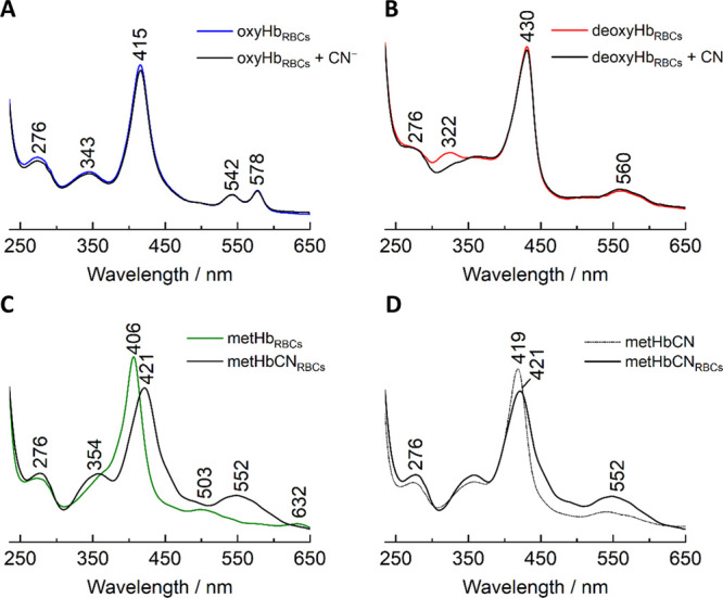
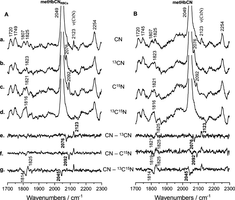
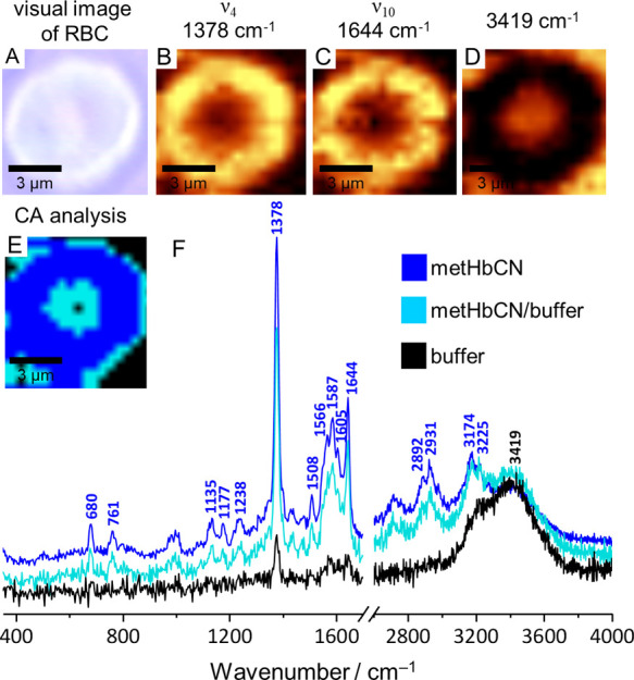
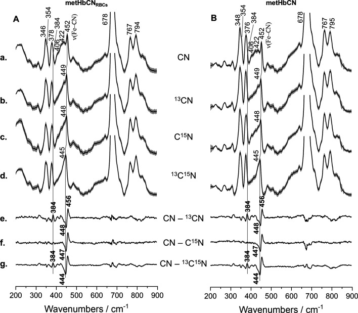
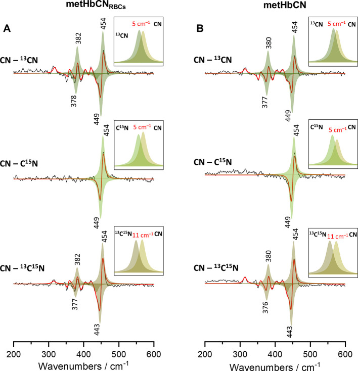
The UV-vis absorption, Raman imaging, and resonance Raman (rR) spectroscopy methods were employed to study cyanohemoglobin (HbCN) adducts inside living functional red blood cells (RBCs). The cyanide ligands are especially optically sensitive probes of the active site environment of heme proteins. The rR studies of HbCN and its isotopic analogues (CN, CN, and CN), as well as a careful deconvolution of spectral data, revealed that the ν(Fe-CN) stretching, δ(Fe-CN) bending, and ν(C≡N) stretching modes occur at 454, 382, and 2123 cm, respectively. Interestingly, while the ν(Fe-CN) modes exhibit the same frequencies in both the isolated and RBC-enclosed hemoglobin molecules, small frequency differences are observed in the δ(Fe-CN) bending modes and the values of their isotopic shifts. These studies show that even though the overall tilted conformation of the Fe-C≡N fragment in the isolated HbCN is preserved in the HbCN enclosed within living cells, there is a small difference in the degree of distortion of the Fe-C≡N fragment. The slight changes in the ligand geometry can be reasonably attributed to the high ordering and tight packing of Hb molecules inside RBCs.
采用紫外可见吸收光谱、拉曼成像和共振拉曼光谱方法研究了活的功能性红细胞(RBC)内的氰化血红蛋白(HbCN)加合物。氰化物配体是血红素蛋白活性位点环境的特别灵敏的光学探针。对HbCN及其同位素类似物(¹²C¹⁴N、¹³C¹⁴N和¹⁵N¹²C)的共振拉曼研究以及对光谱数据的仔细去卷积分析表明,ν(Fe-CN)伸缩振动、δ(Fe-CN)弯曲振动和ν(C≡N)伸缩振动模式分别出现在454、382和2123 cm⁻¹处。有趣的是,虽然ν(Fe-CN)模式在分离的血红蛋白分子和红细胞内的血红蛋白分子中表现出相同的频率,但在δ(Fe-CN)弯曲振动模式及其同位素位移值中观察到了小的频率差异。这些研究表明,尽管分离的HbCN中Fe-C≡N片段的整体倾斜构象在活细胞内的HbCN中得以保留,但Fe-C≡N片段的扭曲程度存在微小差异。配体几何结构的轻微变化可以合理地归因于红细胞内Hb分子的高度有序排列和紧密堆积。