Platzman Ilia, Muth Christine Anna, Lee-Thedieck Cornelia, Pallarola Diego, Atanasova Ralitsa, Louban Ilia, Altrock Eva, Spatz Joachim P
Department of New Materials and Biosystems, Max Planck Institute for Intelligent Systems Heisenbergstr. 3, Stuttgart 70569, Germany & Department of Biophysical Chemistry, University of Heidelberg, Heidelberg 69120, Germany.
Karlsruhe Institute of Technology (KIT), Institute of Functional Interfaces, Hermann-von Helmholtz-Platz 1, 76344 Eggenstein-Leopoldshafen, Germany.
RSC Adv. 2013 Aug 28;3(32):13293-13303. doi: 10.1039/C3RA41579A. Epub 2013 Jun 21.
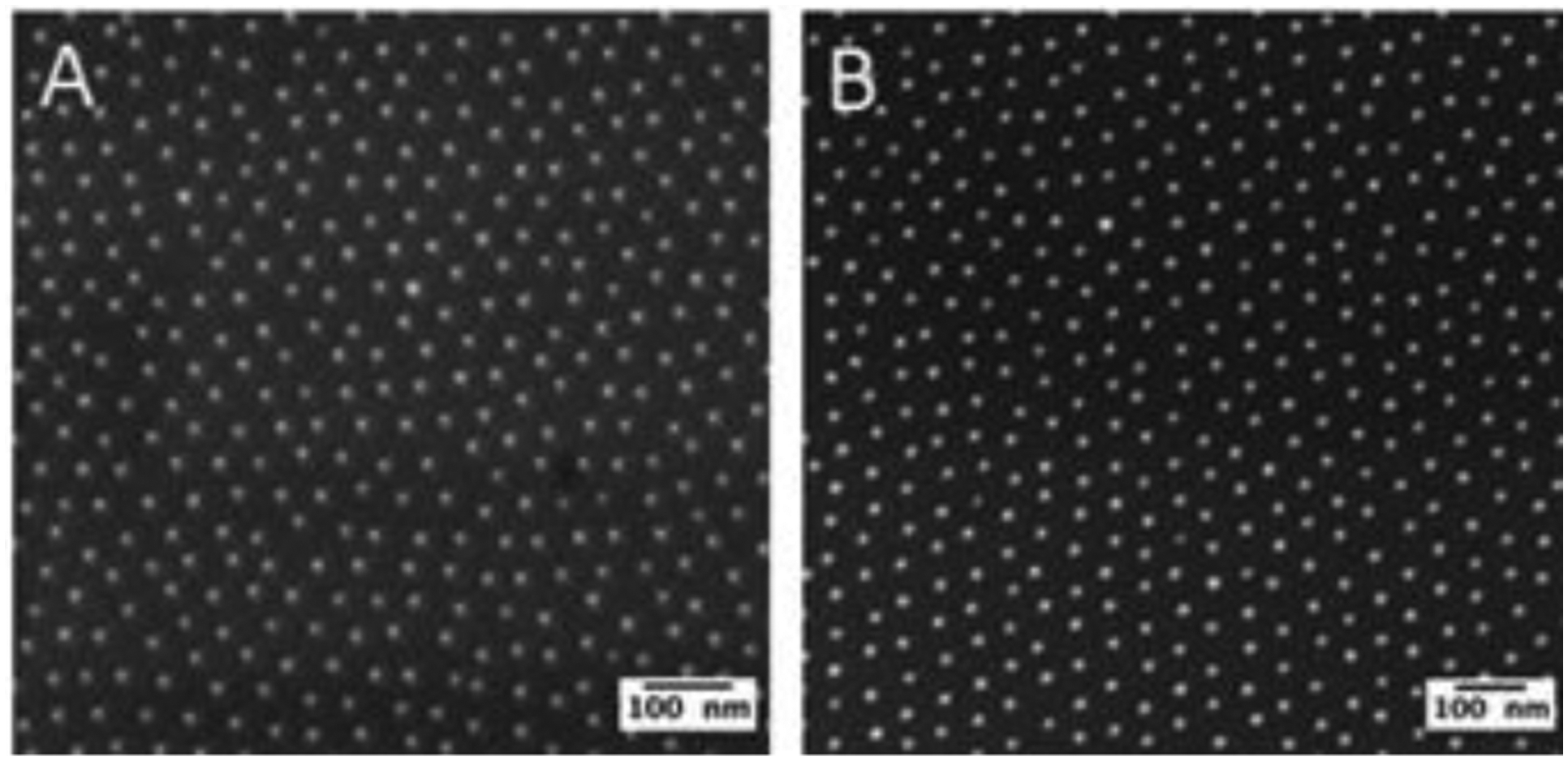
Due to their ability to confer key functions of the native extracellular matrix (ECM) poly(ethylene glycol) (PEG)-based and PEG-modified materials have been extensively used as biocompatible and biofunctionalized substrate systems to study the influence of environmental parameters on cell adhesion Given wide-ranging recent evidence that ECM compliance influences a variety of cell functions, the detailed determination and characterization of the specific PEG surface characteristics including topography, stiffness and chemistry is required. Here, we studied two frequently used bio-active interfaces - PEG-based and PEG-modified surfaces - to elucidate the differences between the physical surface properties, which cells can sense and respond to. For this purpose, two sets of surfaces were synthesized: the first set consisted of nanopatterned glass surfaces containing RGD-functionalized gold nanoparticles surrounded by a passivated PEG-silane layer and the second set consisted of PEG-diacrylate (PEG-DA) hydrogels decorated with RGD-functionalized gold nanoparticlesAlthough the two sets of nanostructured materials compared here were highly similar in terms of density and geometrical distribution of the presented bio-ligands as well as in terms of mechanical bulk properties, the topography and mechanical properties of the surfaces were found to be substantially different and are described in detail. In comparison to very stiff and ultrasmooth surface properties of the PEG-passivated glasses, the mechanical properties of PEG-DA surfaces in the biologically relevant stiffness range, together with the increased surface roughness at micro- and nanoscale levels have the potential to affect cell behavior. This potential was verified by studying the adhesive behavior of hematopoietic KG-1a and rat embryonic fibroblast (REF52) cells on both surfaces.
由于聚乙二醇(PEG)基材料和PEG改性材料能够赋予天然细胞外基质(ECM)关键功能,它们已被广泛用作生物相容性和生物功能化的底物系统,以研究环境参数对细胞黏附的影响。鉴于最近大量证据表明ECM的顺应性会影响多种细胞功能,因此需要详细测定和表征包括形貌、硬度和化学性质在内的特定PEG表面特性。在这里,我们研究了两种常用的生物活性界面——PEG基表面和PEG改性表面——以阐明细胞能够感知并做出反应的物理表面性质之间的差异。为此,合成了两组表面:第一组由纳米图案化的玻璃表面组成,其中含有RGD功能化的金纳米颗粒,并被钝化的PEG硅烷层包围;第二组由装饰有RGD功能化金纳米颗粒的聚乙二醇二丙烯酸酯(PEG-DA)水凝胶组成。尽管这里比较的两组纳米结构材料在所呈现的生物配体的密度和几何分布以及机械整体性质方面非常相似,但发现表面的形貌和机械性质有很大不同,并将进行详细描述。与PEG钝化玻璃非常坚硬和超光滑的表面性质相比,PEG-DA表面在生物学相关硬度范围内的机械性质,以及在微米和纳米尺度上增加的表面粗糙度,有可能影响细胞行为。通过研究造血KG-1a细胞和大鼠胚胎成纤维细胞(REF52)在这两种表面上的黏附行为,验证了这种可能性。