Department of Orthopaedics, University of Utah, Salt Lake City, 84108, USA.
Department of Orthopaedics, Kantonsspital Baselland, 4410, Liestal, Switzerland.
Sci Rep. 2021 Apr 1;11(1):7314. doi: 10.1038/s41598-021-86567-7.
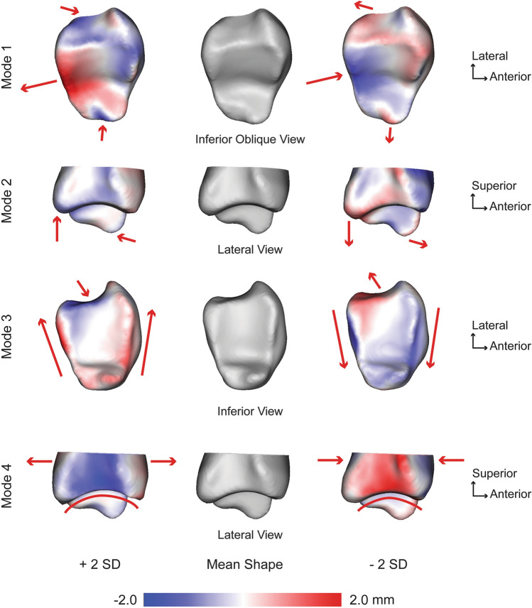
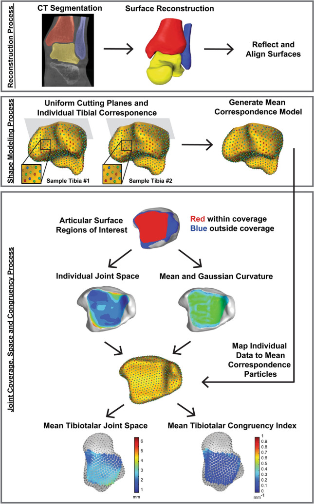
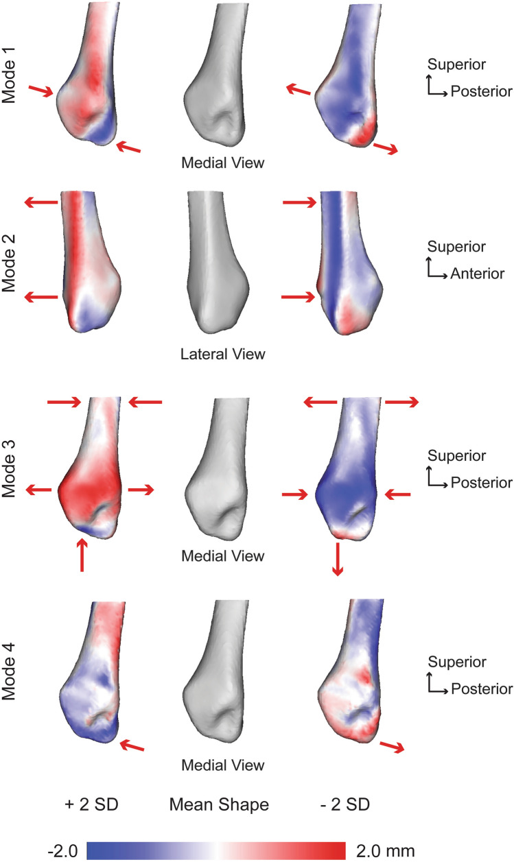
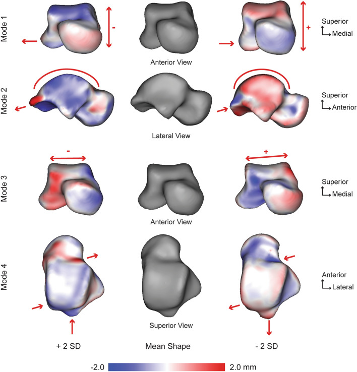
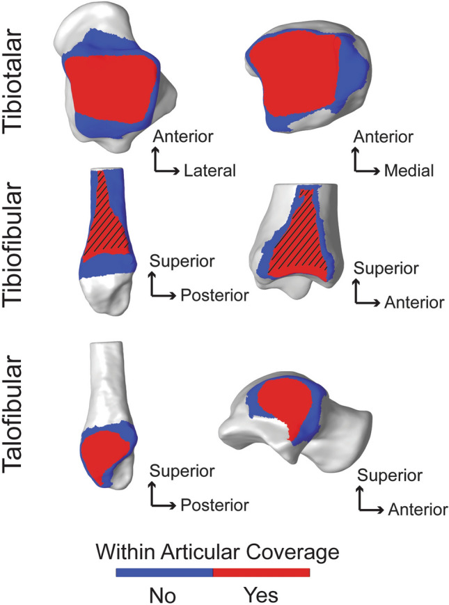
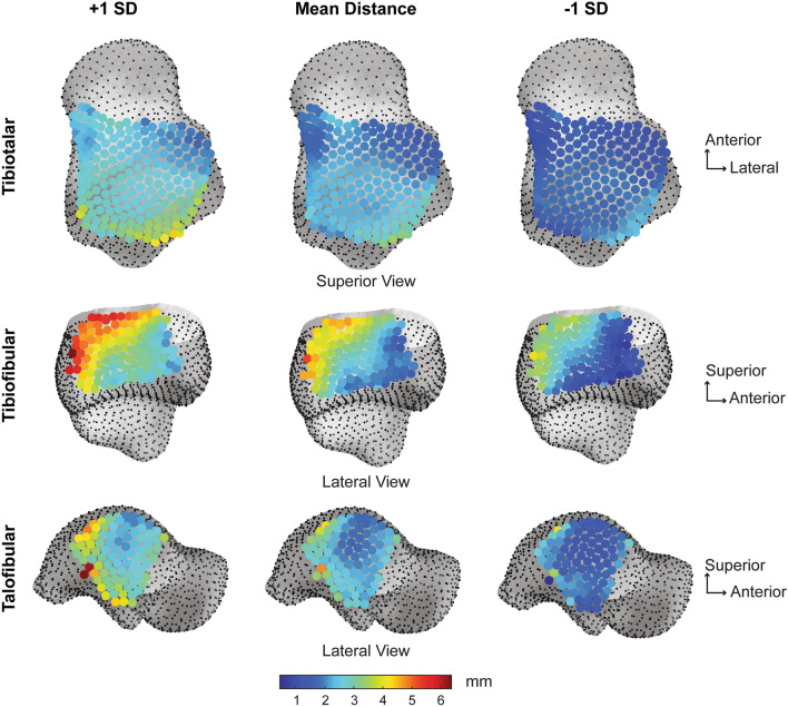
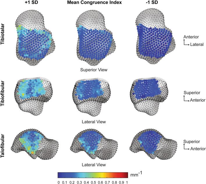
Historically, conventional radiographs have been the primary tool to morphometrically evaluate the talocrural joint, which is comprised of the distal tibia, distal fibula, and proximal talus. More recently, high-resolution volumetric imaging, including computed tomography (CT), has enabled the generation of three-dimensional (3D) reconstructions of the talocrural joint. Weightbearing cone-beam CT (WBCT) technology provides additional benefit to assess 3D spatial relationships and joint congruency while the patient is load bearing. In this study we applied statistical shape modeling, a computational morphometrics technique, to objectively quantify anatomical variation, joint level coverage, joint space distance, and congruency at the talocrural joint. Shape models were developed from segmented WBCT images and included the distal tibia, distal fibula, and full talus. Key anatomical variation across subjects included the fibular notch on the tibia, talar trochlea sagittal plane rate of curvature, tibial plafond curvature with medial malleolus prominence, and changes in the fibular shaft diameter. The shape analysis also revealed a highly congruent talocrural joint with minimal inter-individual morphometric differences at the articular regions. These data are helpful to improve understanding of ankle joint pathologies and to guide refinement of operative treatments.
从历史上看,传统的 X 光片一直是定量评估距下关节的主要工具,距下关节由胫骨远端、腓骨远端和距骨近端组成。最近,高分辨率容积成像,包括计算机断层扫描(CT),已经能够生成距下关节的三维(3D)重建。负重锥形束 CT(WBCT)技术在患者负重时提供了评估 3D 空间关系和关节一致性的额外益处。在这项研究中,我们应用了统计形状建模,这是一种计算形态计量学技术,客观地量化了距下关节的解剖变异、关节水平覆盖范围、关节间隙距离和一致性。形状模型是从分割的 WBCT 图像中开发的,包括胫骨远端、腓骨远端和整个距骨。受试者之间的主要解剖变异包括胫骨上的腓骨切迹、距骨滑车矢状面曲率率、内侧踝突出的胫骨平台曲率以及腓骨干直径的变化。形状分析还显示了距下关节高度一致,关节区域的个体间形态计量差异最小。这些数据有助于提高对踝关节疾病的理解,并指导手术治疗的改进。