Centre for Molecular Nanometrology, Department of Pure and Applied Chemistry, Technology and Innovation Centre, University of Strathclyde, 99 George Street, Glasgow G1 1RD, UK.
MRC Institute of Genetics and Molecular Medicine, Edinburgh Cancer Research UK Centre, University of Edinburgh, Western General Hospital, Crewe Road South, Edinburgh EH4 2XU, UK.
Anal Chem. 2021 Apr 13;93(14):5862-5871. doi: 10.1021/acs.analchem.1c00188. Epub 2021 Apr 2.
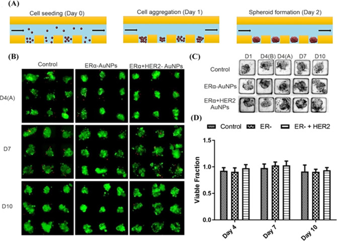
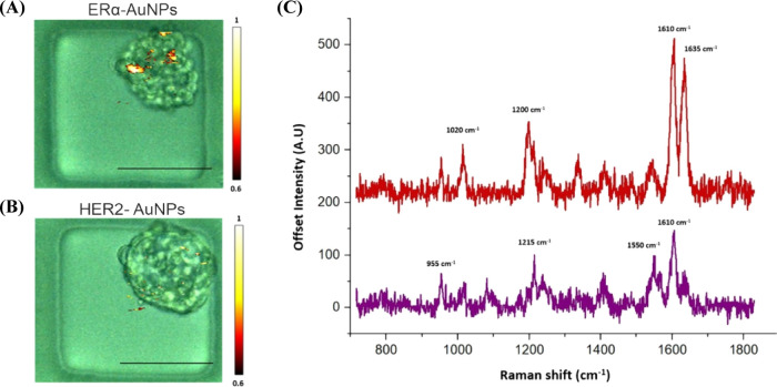
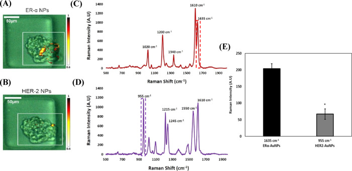
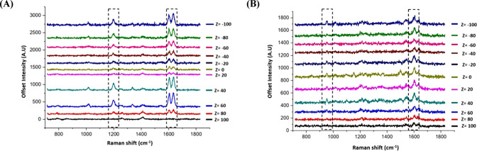
Breast cancer is one of the leading causes of cancer death in women. Novel in vitro tools that integrate three-dimensional (3D) tumor models with highly sensitive chemical reporters can provide useful information to aid biological characterization of cancer phenotype and understanding of drug activity. The combination of surface-enhanced Raman scattering (SERS) techniques with microfluidic technologies offers new opportunities for highly selective, specific, and multiplexed nanoparticle-based assays. Here, we explored the use of functionalized nanoparticles for the detection of estrogen receptor alpha (ERα) expression in a 3D tumor model, using the ERα-positive human breast cancer cell line MCF-7. This approach was used to compare targeted versus nontargeted nanoparticle interactions with the tumor model to better understand whether targeted nanotags are required to efficiently target ERα. Mixtures of targeted anti-ERα antibody-functionalized nanotags (ERα-AuNPs) and nontargeted (against ERα) anti-human epidermal growth factor receptor 2 (HER2) antibody-functionalized nanotags (HER2-AuNPs), with different Raman reporters with a similar SERS signal intensity, were incubated with MCF-7 spheroids in microfluidic devices and spectroscopically analyzed using SERS. MCF-7 cells express high levels of ERα and no detectable levels of HER2. 2D and 3D SERS measurements confirmed the strong targeting effect of ERα-AuNP nanotags to the MCF-7 spheroids in contrast to HER2-AuNPs (63% signal reduction). Moreover, 3D SERS measurements confirmed the differentiation between the targeted and the nontargeted nanotags. Finally, we demonstrated how nanotag uptake by MCF-7 spheroids was affected by the drug fulvestrant, the first-in-class approved selective estrogen receptor degrader (SERD). These results illustrate the potential of using SERS and microfluidics as a powerful in vitro platform for the characterization of 3D tumor models and the investigation of SERD activity.
乳腺癌是导致女性癌症死亡的主要原因之一。新型的体外工具将三维(3D)肿瘤模型与高灵敏度化学报告器相结合,可以提供有用的信息,以辅助癌症表型的生物学特征描述和对药物活性的理解。表面增强拉曼散射(SERS)技术与微流控技术的结合为基于纳米粒子的高度选择性、特异性和多重分析提供了新的机会。在这里,我们探索了使用功能化纳米粒子检测 3D 肿瘤模型中雌激素受体 alpha(ERα)的表达,使用 ERα 阳性的人乳腺癌细胞系 MCF-7。这种方法用于比较靶向与非靶向纳米粒子与肿瘤模型的相互作用,以更好地理解是否需要靶向纳米标签来有效地靶向 ERα。靶向抗 ERα 抗体功能化纳米标签(ERα-AuNPs)和非靶向(针对 ERα)抗人表皮生长因子受体 2(HER2)抗体功能化纳米标签(HER2-AuNPs)的混合物,具有类似 SERS 信号强度的不同 Raman 报告器,与 MCF-7 球体在微流控装置中孵育,并使用 SERS 进行光谱分析。MCF-7 细胞表达高水平的 ERα,且无法检测到 HER2。2D 和 3D SERS 测量结果证实,与 HER2-AuNPs(信号减少 63%)相比,ERα-AuNP 纳米标签对 MCF-7 球体具有很强的靶向作用。此外,3D SERS 测量结果证实了靶向和非靶向纳米标签之间的区别。最后,我们展示了 MCF-7 球体对纳米标签的摄取如何受药物氟维司群(批准的首个选择性雌激素受体降解剂(SERD))的影响。这些结果表明,使用 SERS 和微流控技术作为体外平台来描述 3D 肿瘤模型和研究 SERD 活性具有潜力。