• 文献检索
  • 文档翻译
  • 深度研究
  • 学术资讯
  • Suppr Zotero 插件Zotero 插件
  • 邀请有礼
  • 套餐&价格
  • 历史记录
应用&插件
Suppr Zotero 插件Zotero 插件浏览器插件Mac 客户端Windows 客户端微信小程序
定价
高级版会员购买积分包购买API积分包
服务
文献检索文档翻译深度研究API 文档MCP 服务
关于我们
关于 Suppr公司介绍联系我们用户协议隐私条款
关注我们

Suppr 超能文献

核心技术专利:CN118964589B侵权必究
粤ICP备2023148730 号-1Suppr @ 2026

文献检索

告别复杂PubMed语法,用中文像聊天一样搜索,搜遍4000万医学文献。AI智能推荐,让科研检索更轻松。

立即免费搜索

文件翻译

保留排版,准确专业,支持PDF/Word/PPT等文件格式,支持 12+语言互译。

免费翻译文档

深度研究

AI帮你快速写综述,25分钟生成高质量综述,智能提取关键信息,辅助科研写作。

立即免费体验

从18F-FDG-PET/CT提取的传统PET参数和放射组学特征对肺神经内分泌肿瘤组织学亚型分类及特征描述的诊断价值

Diagnostic Value of Conventional PET Parameters and Radiomic Features Extracted from 18F-FDG-PET/CT for Histologic Subtype Classification and Characterization of Lung Neuroendocrine Neoplasms.

作者信息

Thuillier Philippe, Liberini Virginia, Rampado Osvaldo, Gallio Elena, De Santi Bruno, Ceci Francesco, Metovic Jasna, Papotti Mauro, Volante Marco, Molinari Filippo, Deandreis Désirée

机构信息

Nuclear Medicine Unit, Department of Medical Sciences, University of Turin, 10126 Turin, Italy.

Department of Endocrinology, University Hospital of Brest, 29200 Brest, France.

出版信息

Biomedicines. 2021 Mar 10;9(3):281. doi: 10.3390/biomedicines9030281.

DOI:10.3390/biomedicines9030281
PMID:33801987
原文链接:https://pmc.ncbi.nlm.nih.gov/articles/PMC8001140/
Abstract

AIM

To evaluate if conventional Positron emission tomography (PET) parameters and radiomic features (RFs) extracted by 18F-FDG-PET/CT can differentiate among different histological subtypes of lung neuroendocrine neoplasms (Lu-NENs).

METHODS

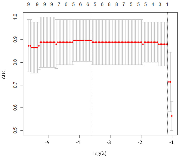
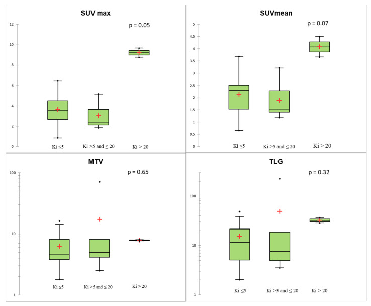
Forty-four naïve-treatment patients on whom 18F-FDG-PET/CT was performed for histologically confirmed Lu-NEN (n = 46) were retrospectively included. Manual segmentation was performed by two operators allowing for extraction of four conventional PET parameters (SUVmax, SUVmean, metabolic tumor volume (MTV), and total lesion glycolysis (TLG)) and 41 RFs. Lu-NENs were classified into two groups: lung neuroendocrine tumors (Lu-NETs) vs. lung neuroendocrine carcinomas (Lu-NECs). Lu-NETs were classified according to histological subtypes (typical (TC)/atypical carcinoid (AC)), Ki67-level, and TNM staging. The least absolute shrink age and selection operator (LASSO) method was used to select the most predictive RFs for classification and Pearson correlation analysis was performed between conventional PET parameters and selected RFs.

RESULTS

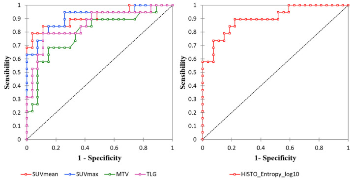
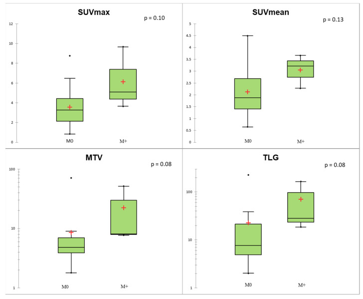
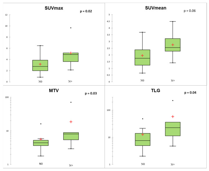
PET parameters, in particular, SUVmax (area under the curve (AUC) = 0.91; cut-off = 5.16) were higher in Lu-NECs vs. Lu-NETs ( < 0.001). Among RFs, HISTO_Entropy_log10 was the most predictive (AUC = 0.90), but correlated with SUVmax/SUVmean (r = 0.95/r = 0.94, respectively). No statistical differences were found between conventional PET parameters and RFs ( > 0.05) and TC vs. AC classification. Conventional PET parameters were correlated with N+ status in Lu-NETs.

CONCLUSION

In our study, conventional PET parameters were able to distinguish Lu-NECs from Lu-NETs, but not TC from AC. RFs did not provide additional information.

摘要

目的

评估常规正电子发射断层扫描(PET)参数以及通过18F-FDG-PET/CT提取的放射组学特征(RFs)能否区分肺神经内分泌肿瘤(Lu-NENs)的不同组织学亚型。

方法

回顾性纳入44例初治患者,这些患者因组织学确诊为Lu-NEN(n = 46)而接受了18F-FDG-PET/CT检查。由两名操作人员进行手动分割,以提取四个常规PET参数(最大标准摄取值(SUVmax)、平均标准摄取值(SUVmean)、代谢肿瘤体积(MTV)和总病变糖酵解(TLG))以及41个RFs。Lu-NENs分为两组:肺神经内分泌肿瘤(Lu-NETs)与肺神经内分泌癌(Lu-NECs)。Lu-NETs根据组织学亚型(典型类癌(TC)/非典型类癌(AC))、Ki67水平和TNM分期进行分类。使用最小绝对收缩和选择算子(LASSO)方法选择用于分类的最具预测性的RFs,并对常规PET参数与所选RFs进行Pearson相关性分析。

结果

PET参数,尤其是SUVmax(曲线下面积(AUC)= 0.91;截断值 = 5.16)在Lu-NECs中高于Lu-NETs(< 0.001)。在RFs中,HISTO_Entropy_log10最具预测性(AUC = 0.90),但与SUVmax/SUVmean相关(分别为r = 0.95/r = 0.94)。常规PET参数与RFs之间(> 0.05)以及TC与AC分类之间未发现统计学差异。常规PET参数与Lu-NETs中的N+状态相关。

结论

在我们的研究中,常规PET参数能够区分Lu-NECs与Lu-NETs,但不能区分TC与AC。RFs未提供额外信息。

https://cdn.ncbi.nlm.nih.gov/pmc/blobs/58eb/8001140/6787de942b18/biomedicines-09-00281-g005.jpg
https://cdn.ncbi.nlm.nih.gov/pmc/blobs/58eb/8001140/02cc35505297/biomedicines-09-00281-g001.jpg
https://cdn.ncbi.nlm.nih.gov/pmc/blobs/58eb/8001140/f74ce45b22b5/biomedicines-09-00281-g002.jpg
https://cdn.ncbi.nlm.nih.gov/pmc/blobs/58eb/8001140/f4211d3afd67/biomedicines-09-00281-g003.jpg
https://cdn.ncbi.nlm.nih.gov/pmc/blobs/58eb/8001140/695029385338/biomedicines-09-00281-g004.jpg
https://cdn.ncbi.nlm.nih.gov/pmc/blobs/58eb/8001140/6787de942b18/biomedicines-09-00281-g005.jpg

相似文献

1
Diagnostic Value of Conventional PET Parameters and Radiomic Features Extracted from 18F-FDG-PET/CT for Histologic Subtype Classification and Characterization of Lung Neuroendocrine Neoplasms.从18F-FDG-PET/CT提取的传统PET参数和放射组学特征对肺神经内分泌肿瘤组织学亚型分类及特征描述的诊断价值
Biomedicines. 2021 Mar 10;9(3):281. doi: 10.3390/biomedicines9030281.
2
Molecular subtype classification of breast cancer using established radiomic signature models based on F-FDG PET/CT images.基于 F-FDG PET/CT 图像的放射组学特征模型对乳腺癌进行分子亚型分类。
Front Biosci (Landmark Ed). 2021 Aug 30;26(9):475-484. doi: 10.52586/4960.
3
Predictive Power of a Radiomic Signature Based on F-FDG PET/CT Images for EGFR Mutational Status in NSCLC.基于F-FDG PET/CT图像的放射组学特征对非小细胞肺癌EGFR突变状态的预测能力
Front Oncol. 2019 Oct 15;9:1062. doi: 10.3389/fonc.2019.01062. eCollection 2019.
4
Dual Tracer 68Ga-DOTATOC and 18F-FDG PET Improve Preoperative Evaluation of Aggressiveness in Resectable Pancreatic Neuroendocrine Neoplasms.双示踪剂68Ga-DOTATOC和18F-FDG PET改善可切除胰腺神经内分泌肿瘤侵袭性的术前评估。
Diagnostics (Basel). 2021 Jan 28;11(2):192. doi: 10.3390/diagnostics11020192.
5
Assessment of Mediastinal Tumors Using SUV and Volumetric Parameters on FDG-PET/CT.利用 FDG-PET/CT 上的 SUV 和体积参数评估纵隔肿瘤
Asia Ocean J Nucl Med Biol. 2017 Winter;5(1):22-29. doi: 10.22038/aojnmb.2016.7996.
6
Prognostic Value of Metabolic and Volumetric Parameters of Preoperative FDG-PET/CT in Patients With Resectable Pancreatic Cancer.术前FDG-PET/CT代谢和容积参数对可切除胰腺癌患者的预后价值
Medicine (Baltimore). 2016 May;95(19):e3686. doi: 10.1097/MD.0000000000003686.
7
F-FDG PET/CT in lung adenosquamous carcinoma and its correlation with clinicopathological features and prognosis.18F-FDG PET/CT 显像在肺鳞腺癌中的表现及其与临床病理特征和预后的关系。
Ann Nucl Med. 2020 May;34(5):314-321. doi: 10.1007/s12149-020-01450-1. Epub 2020 Feb 22.
8
The Correlation Between [Ga]DOTATATE PET/CT and Cell Proliferation in Patients With GEP-NENs.胃泌素瘤神经内分泌肿瘤患者 [Ga]DOTATATE PET/CT 与细胞增殖的相关性。
Mol Imaging Biol. 2019 Oct;21(5):984-990. doi: 10.1007/s11307-019-01328-3.
9
The clinical value of texture analysis of dual-time-point F-FDG-PET/CT imaging to differentiate between F-FDG-avid benign and malignant pulmonary lesions.双时相 F-FDG-PET/CT 纹理分析在鉴别 F-FDG 摄取的肺良恶性病变中的临床价值。
Eur Radiol. 2020 Mar;30(3):1759-1769. doi: 10.1007/s00330-019-06463-7. Epub 2019 Nov 14.
10
F-FDG Pet Parameters and Radiomics Features Analysis in Advanced Nsclc Treated with Immunotherapy as Predictors of Therapy Response and Survival.以免疫疗法治疗的晚期非小细胞肺癌中F-FDG Pet参数与放射组学特征分析作为治疗反应和生存的预测指标
Cancers (Basel). 2020 May 5;12(5):1163. doi: 10.3390/cancers12051163.

引用本文的文献

1
Application of Radiomics for Differentiating Lung Neuroendocrine Neoplasms.放射组学在鉴别肺神经内分泌肿瘤中的应用
Diagnostics (Basel). 2025 Mar 31;15(7):874. doi: 10.3390/diagnostics15070874.
2
Applications of Artificial Intelligence and Radiomics in Molecular Hybrid Imaging and Theragnostics for Neuro-Endocrine Neoplasms (NENs).人工智能与放射组学在神经内分泌肿瘤(NENs)分子杂交成像及诊疗中的应用
Life (Basel). 2023 Jul 28;13(8):1647. doi: 10.3390/life13081647.
3
The diagnostic and prognostic role of combined [F]FDG and [Ga]-DOTA-peptides PET/CT in primary pulmonary carcinoids: a multicentric experience.

本文引用的文献

1
Lung carcinoids with high proliferative activity: Further support for the identification of a new tumor category in the classification of lung neuroendocrine neoplasms.具有高增殖活性的肺类癌:进一步支持在肺神经内分泌肿瘤分类中识别一种新肿瘤类别的观点。
Lung Cancer. 2020 Oct;148:149-158. doi: 10.1016/j.lungcan.2020.08.001. Epub 2020 Aug 8.
2
Histologic subtype classification of non-small cell lung cancer using PET/CT images.使用 PET/CT 图像对非小细胞肺癌进行组织学亚型分类。
Eur J Nucl Med Mol Imaging. 2021 Feb;48(2):350-360. doi: 10.1007/s00259-020-04771-5. Epub 2020 Aug 10.
3
Theranostic implications of molecular imaging phenotype of well-differentiated pulmonary carcinoid based on Ga-DOTATATE PET/CT and F-FDG PET/CT.
联合 [F]FDG 和 [Ga]-DOTA-肽 PET/CT 在原发性肺类癌中的诊断和预后作用:一项多中心经验。
Eur Radiol. 2023 Jun;33(6):4167-4177. doi: 10.1007/s00330-022-09326-w. Epub 2022 Dec 8.
4
Clinicopathological, Oncogenic, and F-FDG PET/CT Features of Primary Pulmonary Carcinoid in Resection Specimens.切除标本中原发性肺类癌的临床病理、致癌基因和 F-FDG PET/CT 特征。
Contrast Media Mol Imaging. 2022 Jun 15;2022:1926797. doi: 10.1155/2022/1926797. eCollection 2022.
5
Radiomics in Oncological PET Imaging: A Systematic Review-Part 2, Infradiaphragmatic Cancers, Blood Malignancies, Melanoma and Musculoskeletal Cancers.肿瘤PET成像中的放射组学:系统综述 - 第2部分,膈下癌症、血液恶性肿瘤、黑色素瘤和肌肉骨骼肿瘤
Diagnostics (Basel). 2022 May 27;12(6):1330. doi: 10.3390/diagnostics12061330.
6
Somatostatin Receptor-negative and Fluorodeoxyglucose-positron Emission Tomography-positive Lung Neuroendocrine Tumor G1 Exhibiting Cyclic Cushing's Syndrome.生长抑素受体阴性和氟脱氧葡萄糖正电子发射断层扫描阳性肺神经内分泌肿瘤 G1 表现为周期性库欣综合征。
Intern Med. 2022 Dec 15;61(24):3693-3698. doi: 10.2169/internalmedicine.9238-21. Epub 2022 May 31.
7
Radiolabeled Somatostatin Analogues for Diagnosis and Treatment of Neuroendocrine Tumors.用于神经内分泌肿瘤诊断和治疗的放射性标记生长抑素类似物
Cancers (Basel). 2022 Feb 19;14(4):1055. doi: 10.3390/cancers14041055.
8
CT Texture Analysis of Pulmonary Neuroendocrine Tumors-Associations with Tumor Grading and Proliferation.肺神经内分泌肿瘤的CT纹理分析——与肿瘤分级和增殖的相关性
J Clin Med. 2021 Nov 26;10(23):5571. doi: 10.3390/jcm10235571.
9
Evaluating a Machine Learning Tool for the Classification of Pathological Uptake in Whole-Body PSMA-PET-CT Scans.评估一种用于全身PSMA-PET-CT扫描中病理性摄取分类的机器学习工具。
Tomography. 2021 Jul 29;7(3):301-312. doi: 10.3390/tomography7030027.
基于 Ga-DOTATATE PET/CT 和 F-FDG PET/CT 的高分化肺类癌分子影像学表型的治疗意义。
Eur J Nucl Med Mol Imaging. 2021 Jan;48(1):204-216. doi: 10.1007/s00259-020-04915-7. Epub 2020 Jun 22.
4
F-FDG Pet Parameters and Radiomics Features Analysis in Advanced Nsclc Treated with Immunotherapy as Predictors of Therapy Response and Survival.以免疫疗法治疗的晚期非小细胞肺癌中F-FDG Pet参数与放射组学特征分析作为治疗反应和生存的预测指标
Cancers (Basel). 2020 May 5;12(5):1163. doi: 10.3390/cancers12051163.
5
The Image Biomarker Standardization Initiative: Standardized Quantitative Radiomics for High-Throughput Image-based Phenotyping.影像生物标志物标准化倡议:高通量基于影像表型的标准化定量放射组学。
Radiology. 2020 May;295(2):328-338. doi: 10.1148/radiol.2020191145. Epub 2020 Mar 10.
6
Development of a Novel Multiparametric MRI Radiomic Nomogram for Preoperative Evaluation of Early Recurrence in Resectable Pancreatic Cancer.开发一种新型多参数 MRI 放射组学列线图,用于术前评估可切除胰腺癌的早期复发。
J Magn Reson Imaging. 2020 Jul;52(1):231-245. doi: 10.1002/jmri.27024. Epub 2019 Dec 23.
7
Predictive Power of a Radiomic Signature Based on F-FDG PET/CT Images for EGFR Mutational Status in NSCLC.基于F-FDG PET/CT图像的放射组学特征对非小细胞肺癌EGFR突变状态的预测能力
Front Oncol. 2019 Oct 15;9:1062. doi: 10.3389/fonc.2019.01062. eCollection 2019.
8
Characteristics of 252 patients with bronchopulmonary neuroendocrine tumours treated at the Copenhagen NET Centre of Excellence.252 例支气管肺神经内分泌肿瘤患者在哥本哈根神经内分泌肿瘤卓越中心的治疗特征。
Lung Cancer. 2019 Jun;132:141-149. doi: 10.1016/j.lungcan.2019.03.013. Epub 2019 Apr 5.
9
Inter-observer and segmentation method variability of textural analysis in pre-therapeutic FDG PET/CT in head and neck cancer.头颈部癌症治疗前 FDG PET/CT 纹理分析中观察者间和分割方法的可变性。
PLoS One. 2019 Mar 28;14(3):e0214299. doi: 10.1371/journal.pone.0214299. eCollection 2019.
10
The utility of 18F-FDG and 68Ga-DOTA-Peptide PET/CT in the evaluation of primary pulmonary carcinoid: A systematic review and meta-analysis.18F-FDG和68Ga-DOTA-肽PET/CT在原发性肺类癌评估中的应用:一项系统评价和荟萃分析
Medicine (Baltimore). 2019 Mar;98(10):e14769. doi: 10.1097/MD.0000000000014769.