Ko Tsung-Shine, Liu Han-Yuan, Shieh Jiann, Shieh De, Chen Szu-Hung, Chen Yen-Lun, Lin En-Ting
Department of Electronic Engineering, National Changhua University of Education, No. 2, Shi-Da Road, Changhua 50074, Taiwan.
Department of Materials Science and Engineering, National United University, No. 2, Lianda, Miaoli 36063, Taiwan.
Nanomaterials (Basel). 2021 Mar 15;11(3):733. doi: 10.3390/nano11030733.
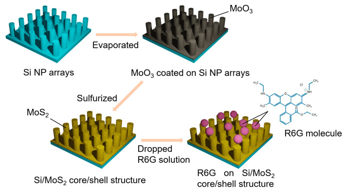
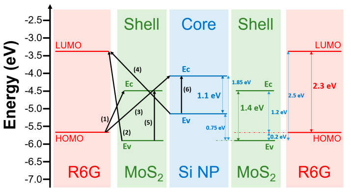
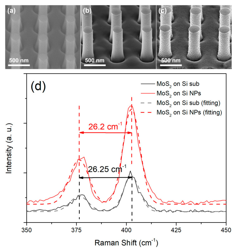
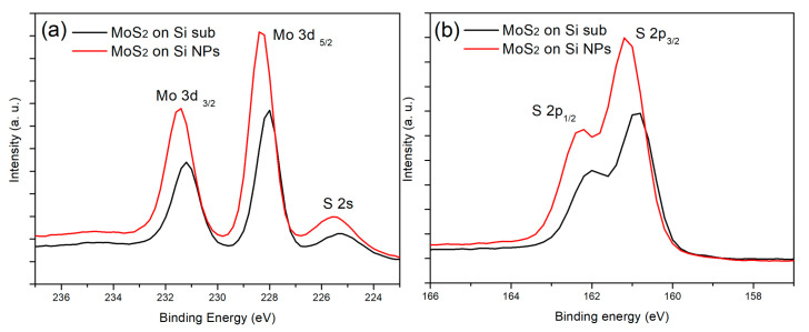
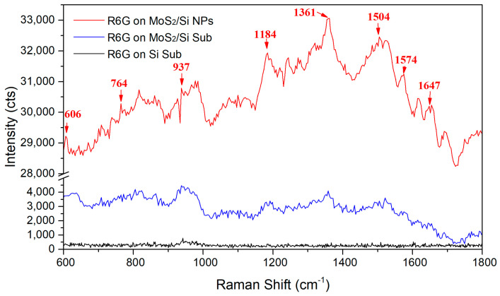
Two-dimensional layered material Molybdenum disulfide (MoS) exhibits a flat surface without dangling bonds and is expected to be a suitable surface-enhanced Raman scattering (SERS) substrate for the detection of organic molecules. However, further fabrication of nanostructures for enhancement of SERS is necessary because of the low detection efficiency of MoS. In this paper, period-distribution Si/MoS core/shell nanopillar (NP) arrays were fabricated for SERS. The MoS thin films were formed on the surface of Si NPs by sulfurizing the MoO thin films coated on the Si NP arrays. Scanning electron microscopy, Raman spectroscopy, and X-ray photoelectron spectroscopy were performed to characterize Si/MoS core-shell nanostructure. In comparison with a bare Si substrate and MoS thin film, the use of Si/MoS core-shell NP arrays as SERS substrates enhances the intensity of each SERS signal peak for Rhodamine 6G (R6G) molecules, and especially exhibits about 75-fold and 7-fold enhancements in the 1361 cm peak signal, respectively. We suggest that the Si/MoS core-shell NP arrays with larger area could absorb more R6G molecules and provide larger interfaces between MoS and R6G molecules, leading to higher opportunity of charge transfer process and exciton transitions. Therefore, the Si/MoS core/shell NP arrays could effectively enhance SERS signal and serve as excellent SERS substrates in biomedical detection.
二维层状材料二硫化钼(MoS)具有无悬键的平坦表面,有望成为用于检测有机分子的合适表面增强拉曼散射(SERS)基底。然而,由于MoS的检测效率较低,因此需要进一步制备用于增强SERS的纳米结构。本文制备了用于SERS的周期分布Si/MoS核壳纳米柱(NP)阵列。通过硫化涂覆在Si NP阵列上的MoO薄膜,在Si NPs表面形成MoS薄膜。进行了扫描电子显微镜、拉曼光谱和X射线光电子能谱表征Si/MoS核壳纳米结构。与裸Si基底和MoS薄膜相比,使用Si/MoS核壳NP阵列作为SERS基底可增强罗丹明6G(R6G)分子的每个SERS信号峰的强度,并且在1361 cm处的峰信号分别表现出约75倍和7倍的增强。我们认为,具有更大面积的Si/MoS核壳NP阵列可以吸收更多的R6G分子,并在MoS和R6G分子之间提供更大的界面,从而导致电荷转移过程和激子跃迁的更高机会。因此,Si/MoS核/壳NP阵列可以有效地增强SERS信号,并在生物医学检测中用作优异的SERS基底。