Maruyama Kazuichi, Yoneda Kazuhito, Sugita Sunao, Yamamoto Yoshimi, Koike Masato, Peters Christoph, Uchiyama Yasuo, Nishida Kohji
Department of Vision Informatics, Osaka University Graduate School of Medicine, Suita 565-0871, Japan.
Integrated Frontier Research for Medical Science Division, Institute for Open and Transdisciplinary Research Initiatives (OTRI), Osaka University, Osaka 565-0871, Japan.
Antioxidants (Basel). 2021 Mar 15;10(3):456. doi: 10.3390/antiox10030456.
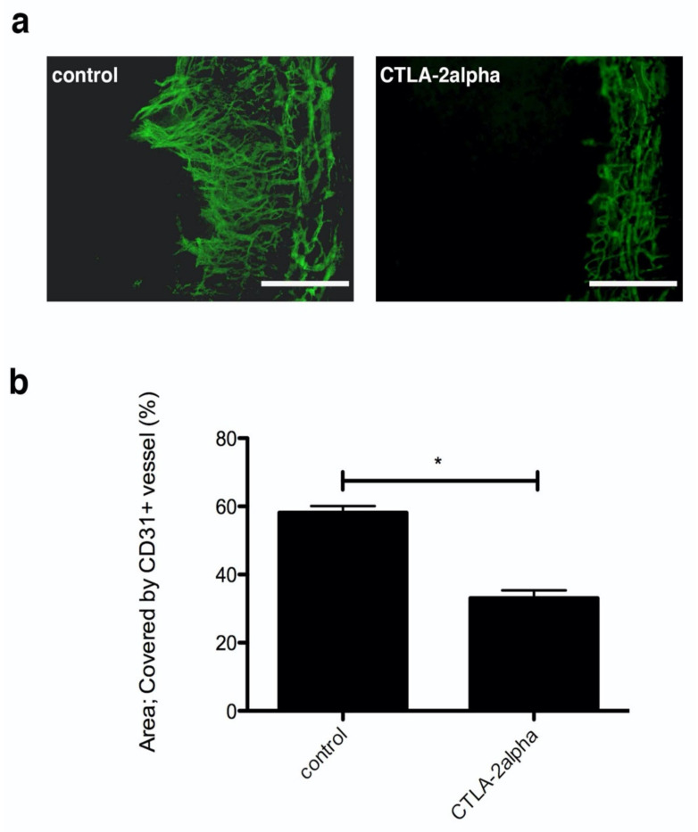
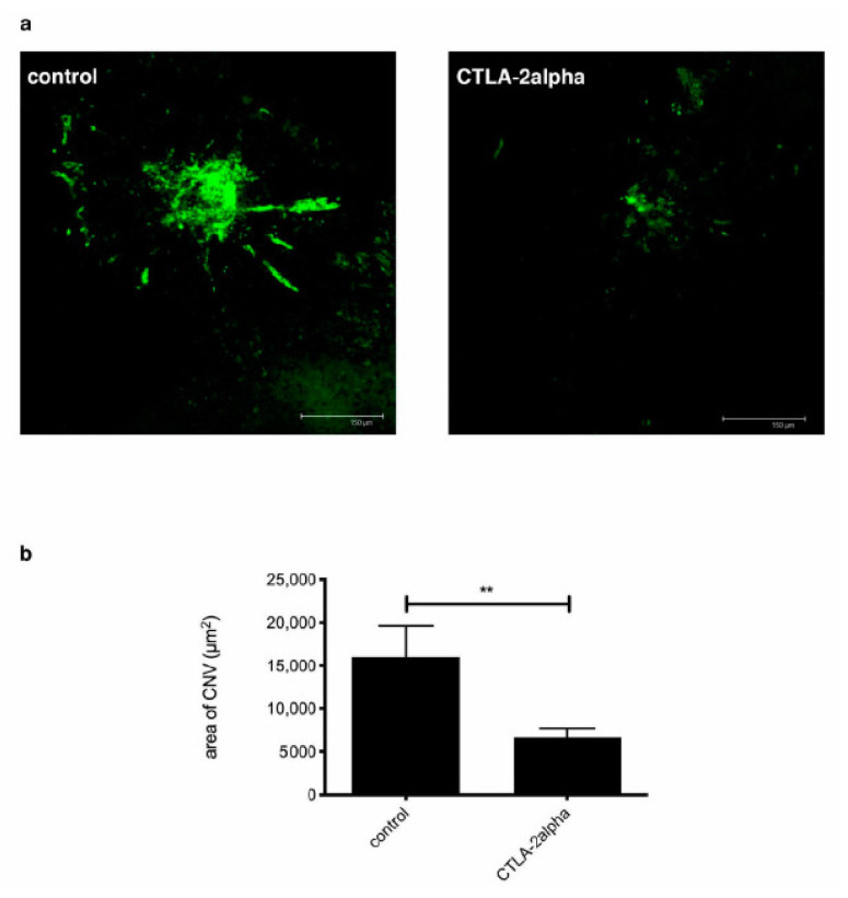
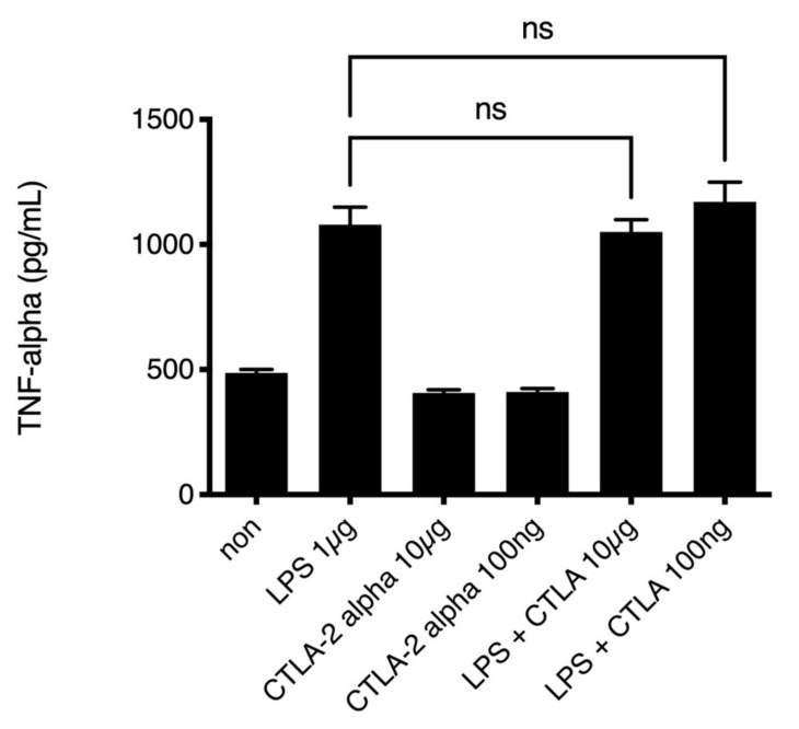
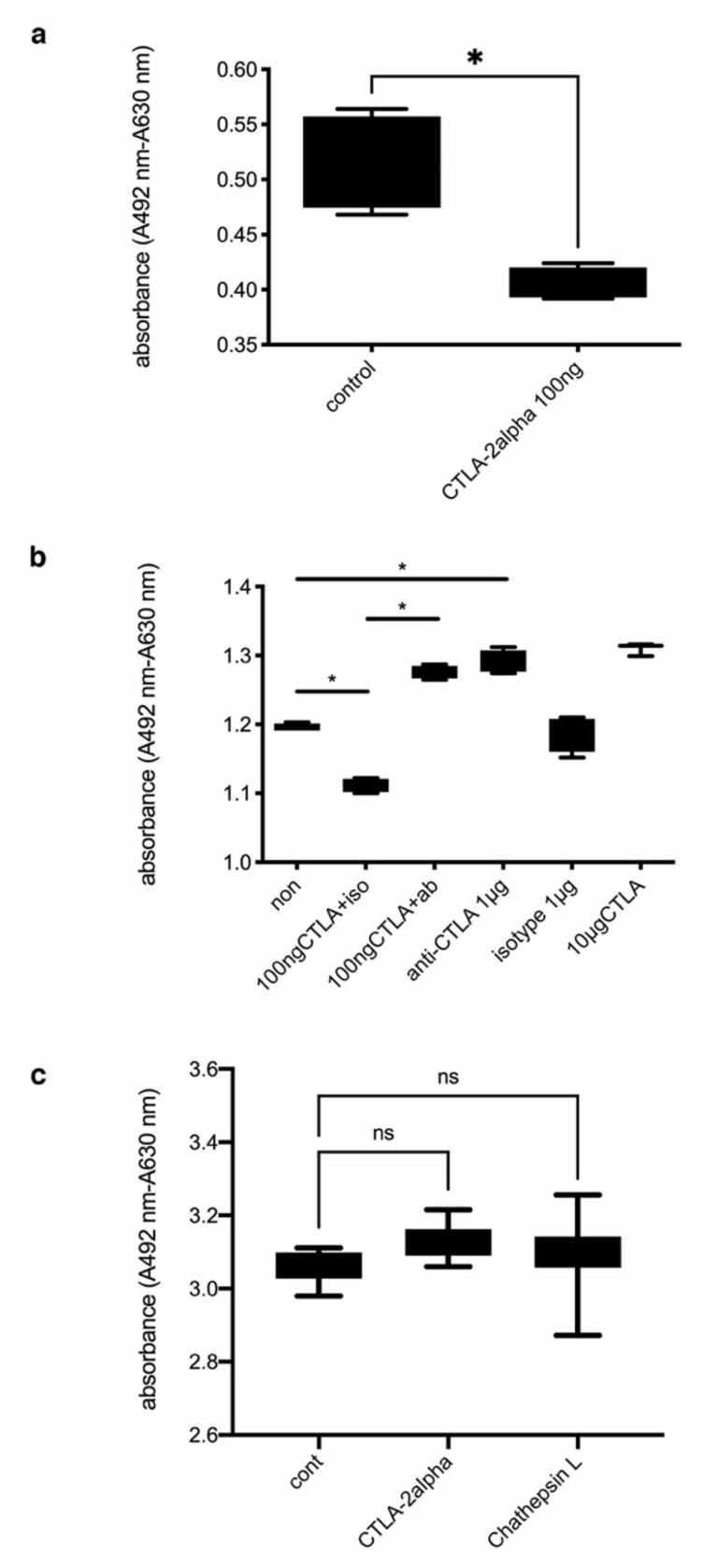
Cytotoxic T lymphocyte antigen-2 (CTLA-2) alpha has been reported to suppress the activities of cathepsin L (Cath L), which is deeply involved in angiogenesis. Therefore, we assessed whether CTLA-2 alpha plays a role in angiogenesis in ocular tissue. To establish models of corneal inflammation and experimental choroidal neovascularization (CNV), male C57BL/6J mice ( = 5) underwent corneal suture placement or laser-induced CNV, respectively. Mice were then injected with recombinant CTLA-2 alpha (1 µg) into the peritoneal cavity at day 0 and every 2 days after operation. In vitro experiments were performed to assess the inflammatory response by measuring TNF-alpha secretion in peritoneal cavity exudate cells (PECs) or the proliferation of mouse vascular endothelial cells (mVECs). CTLA-2 alpha treatment dramatically suppressed corneal angiogenesis, as well as laser-induced CNV. Moreover, CTLA-2 alpha inhibited the proliferation of mVECs in vitro, while CTLA-2 alpha abolishment was able to rescue proliferation. However, CTLA-2 alpha could not suppress cytokine secretion from inflammatory cells such as PECs. In summary, CTLA-2 alpha was able to suppress angiogenesis by suppressing endothelial cell proliferation. Further studies are needed to investigate its usefulness as a new antiangiogenic treatment for a variety of conditions, including age-related macular degeneration.
据报道,细胞毒性T淋巴细胞抗原-2(CTLA-2)α可抑制组织蛋白酶L(组织蛋白酶L)的活性,而组织蛋白酶L与血管生成密切相关。因此,我们评估了CTLA-2α在眼组织血管生成中是否发挥作用。为建立角膜炎症和实验性脉络膜新生血管(CNV)模型,分别对雄性C57BL/6J小鼠(n = 5)进行角膜缝线植入或激光诱导的CNV。然后在术后第0天及之后每2天向小鼠腹腔注射重组CTLA-2α(1μg)。通过测量腹腔渗出细胞(PEC)中TNF-α的分泌或小鼠血管内皮细胞(mVEC)的增殖来进行体外实验,以评估炎症反应。CTLA-2α治疗显著抑制了角膜血管生成以及激光诱导的CNV。此外,CTLA-2α在体外抑制了mVEC的增殖,而CTLA-2α的缺失能够挽救增殖。然而,CTLA-2α不能抑制炎症细胞如PEC的细胞因子分泌。总之,CTLA-2α能够通过抑制内皮细胞增殖来抑制血管生成。需要进一步研究以探讨其作为包括年龄相关性黄斑变性在内的多种病症的新型抗血管生成治疗方法的效用。