Institute of Anatomy, First Faculty of Medicine, Charles University, 128 00 Prague, Czech Republic.
Institute of Physiology, CAS, 142 20 Prague, Czech Republic.
Int J Mol Sci. 2021 Mar 1;22(5):2475. doi: 10.3390/ijms22052475.
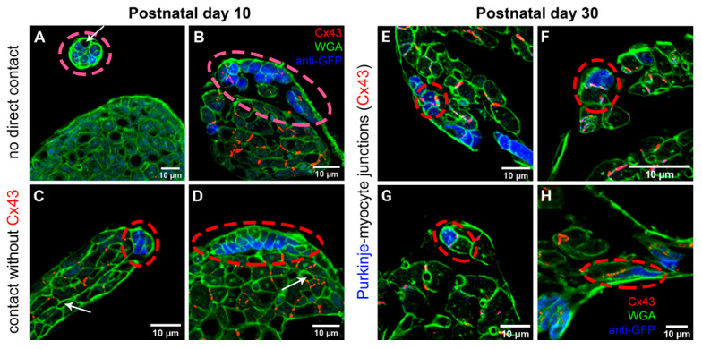
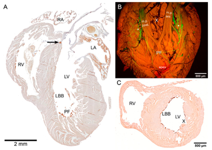
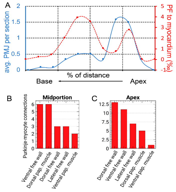
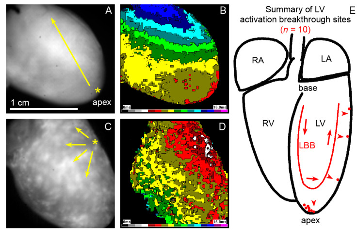
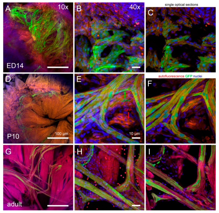
The mammalian ventricular myocardium forms a functional syncytium due to flow of electrical current mediated in part by gap junctions localized within intercalated disks. The connexin (Cx) subunit of gap junctions have direct and indirect roles in conduction of electrical impulse from the cardiac pacemaker via the cardiac conduction system (CCS) to working myocytes. Cx43 is the dominant isoform in these channels. We have studied the distribution of Cx43 junctions between the CCS and working myocytes in a transgenic mouse model, which had the His-Purkinje portion of the CCS labeled with green fluorescence protein. The highest number of such connections was found in a region about one-third of ventricular length above the apex, and it correlated with the peak proportion of Purkinje fibers (PFs) to the ventricular myocardium. At this location, on the septal surface of the left ventricle, the insulated left bundle branch split into the uninsulated network of PFs that continued to the free wall anteriorly and posteriorly. The second peak of PF abundance was present in the ventricular apex. Epicardial activation maps correspondingly placed the site of the first activation in the apical region, while some hearts presented more highly located breakthrough sites. Taken together, these results increase our understanding of the physiological pattern of ventricular activation and its morphological underpinning through detailed CCS anatomy and distribution of its gap junctional coupling to the working myocardium.
哺乳动物的心室心肌由于部分通过位于闰盘内的缝隙连接介导的电流流动而形成功能上的合胞体。缝隙连接的连接蛋白 (Cx) 亚基在心脏起搏冲动通过心脏传导系统 (CCS) 传导至工作心肌的过程中具有直接和间接作用。Cx43 是这些通道中的主要同工型。我们在一种转基因小鼠模型中研究了 CCS 与工作心肌之间的 Cx43 连接的分布,该模型中 CCS 的希氏-浦肯野部分被绿色荧光蛋白标记。在距离心尖约三分之一心室长度的区域发现了最多的这种连接,并且与浦肯野纤维 (PFs) 与心室心肌的比例峰值相关。在这个位置,在左心室的室间隔表面,绝缘的左束支分支成未绝缘的 PF 网络,从前到后继续延伸到游离壁。PF 丰富的第二个峰值出现在心室心尖。心外膜激活图相应地将第一次激活的部位放置在心尖区域,而有些心脏则呈现出更高的突破部位。总的来说,这些结果通过详细的 CCS 解剖结构及其与工作心肌的缝隙连接偶联的分布,增加了我们对心室激活的生理模式及其形态基础的理解。