Olejníčková Veronika, Šaňková Barbora, Sedmera David, Janáček Jiří
Department of Developmental Cardiology, Institute of Physiology of The Czech Academy of Sciences, Prague, Czechia.
First Faculty of Medicine, Charles University, Prague, Czechia.
Front Physiol. 2019 Jan 8;9:1876. doi: 10.3389/fphys.2018.01876. eCollection 2018.
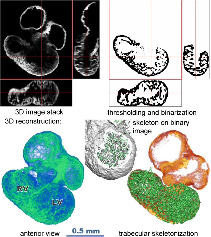
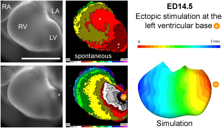
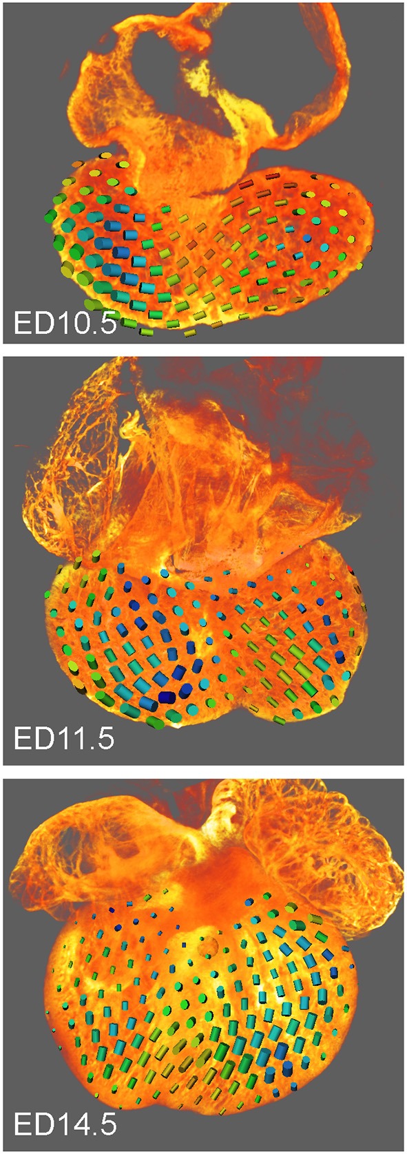
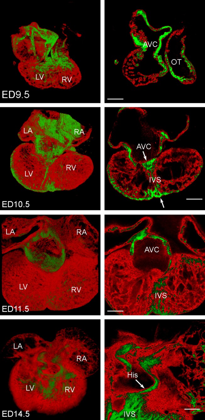
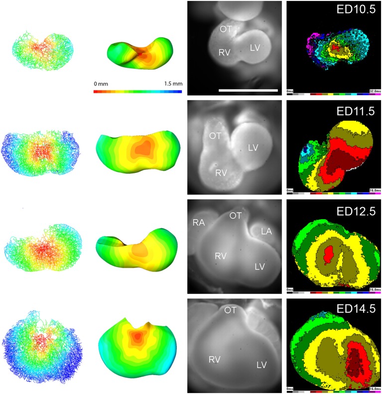
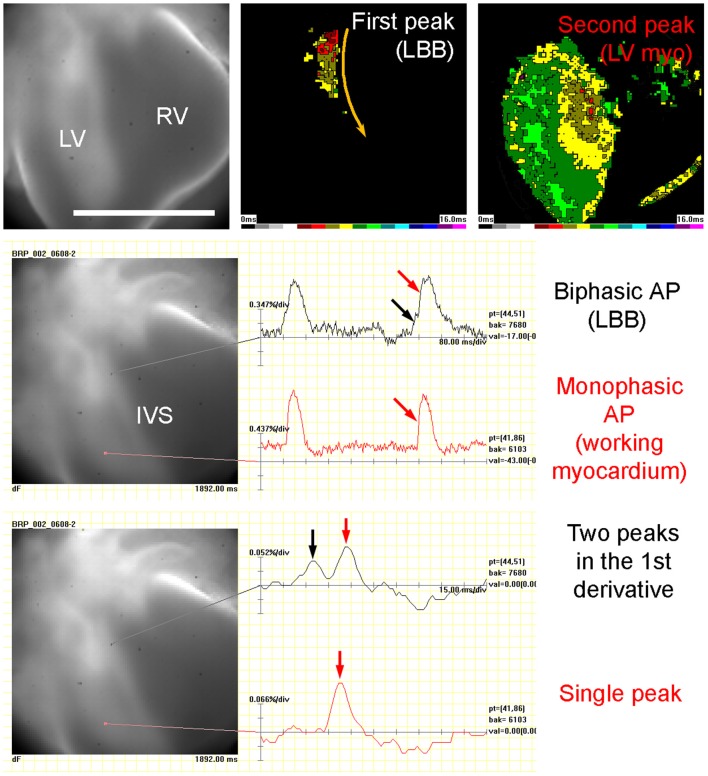
Most embryonic ventricular cardiomyocytes are quite uniform, in contrast to the adult heart, where the specialized ventricular conduction system is molecularly and functionally distinct from the working myocardium. We thus hypothesized that the preferential conduction pathway within the embryonic ventricle could be dictated by trabecular geometry. Mouse embryonic hearts of the Nkx2.5:eGFP strain between ED9.5 and ED14.5 were cleared and imaged whole mount by confocal microscopy, and reconstructed in 3D at 3.4 μm isotropic voxel size. The local orientation of the trabeculae, responsible for the anisotropic spreading of the signal, was characterized using spatially homogenized tensors (3 × 3 matrices) calculated from the trabecular skeleton. Activation maps were simulated assuming constant speed of spreading along the trabeculae. The results were compared with experimentally obtained epicardial activation maps generated by optical mapping with a voltage-sensitive dye. Simulated impulse propagation starting from the top of interventricular septum revealed the first epicardial breakthrough at the interventricular grove, similar to experimentally obtained activation maps. Likewise, ectopic activation from the left ventricular base perpendicular to dominant trabecular orientation resulted in isotropic and slower impulse spreading on the ventricular surface in both simulated and experimental conditions. We conclude that in the embryonic pre-septation heart, the geometry of the A-V connections and trabecular network is sufficient to explain impulse propagation and ventricular activation patterns.
与成体心脏不同,大多数胚胎心室心肌细胞相当均匀,在成体心脏中,专门的心室传导系统在分子和功能上与工作心肌不同。因此,我们推测胚胎心室内的优先传导途径可能由小梁几何形状决定。对ED9.5至ED14.5之间的Nkx2.5:eGFP品系小鼠胚胎心脏进行清除,并通过共聚焦显微镜进行整体成像,以3.4μm各向同性体素大小进行三维重建。利用从小梁骨架计算出的空间均匀张量(3×3矩阵)来表征负责信号各向异性传播的小梁的局部方向。假设沿小梁的传播速度恒定,模拟激活图。将结果与通过电压敏感染料光学映射获得的实验性心外膜激活图进行比较。从室间隔顶部开始的模拟冲动传播显示在心间沟处首次出现心外膜突破,类似于实验获得的激活图。同样,在模拟和实验条件下,从左心室底部垂直于主要小梁方向的异位激活导致冲动在心室表面各向同性且传播较慢。我们得出结论,在胚胎期室间隔形成前的心脏中,房室连接和小梁网络的几何形状足以解释冲动传播和心室激活模式。