Eye Hospital, University Medical Centre Ljubljana, Grablovičeva 46, 1000 Ljubljana, Slovenia.
Clinical Institute of Medical Genetics, University Medical Centre Ljubljana, Šlajmerjeva ulica 4, 1000 Ljubljana, Slovenia.
Genes (Basel). 2021 Mar 29;12(4):499. doi: 10.3390/genes12040499.
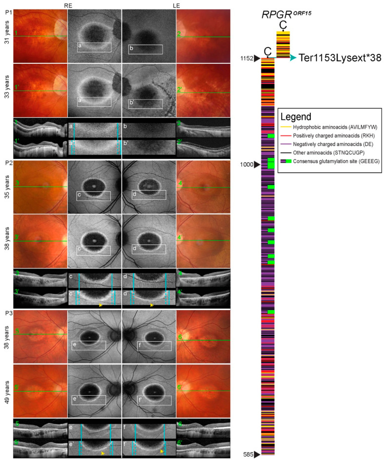
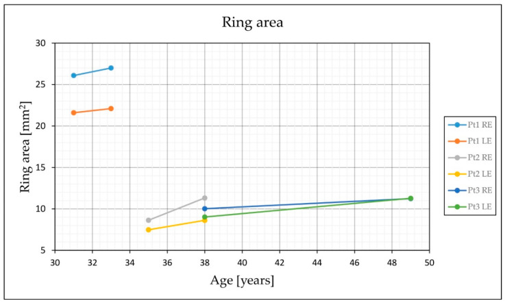
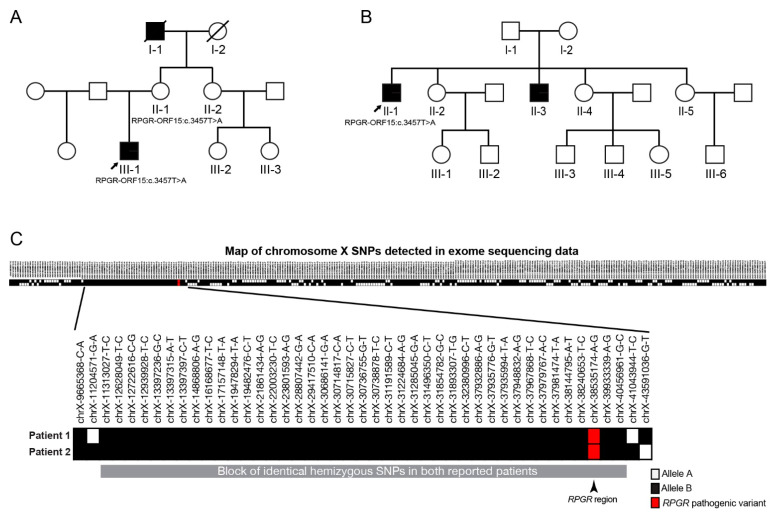
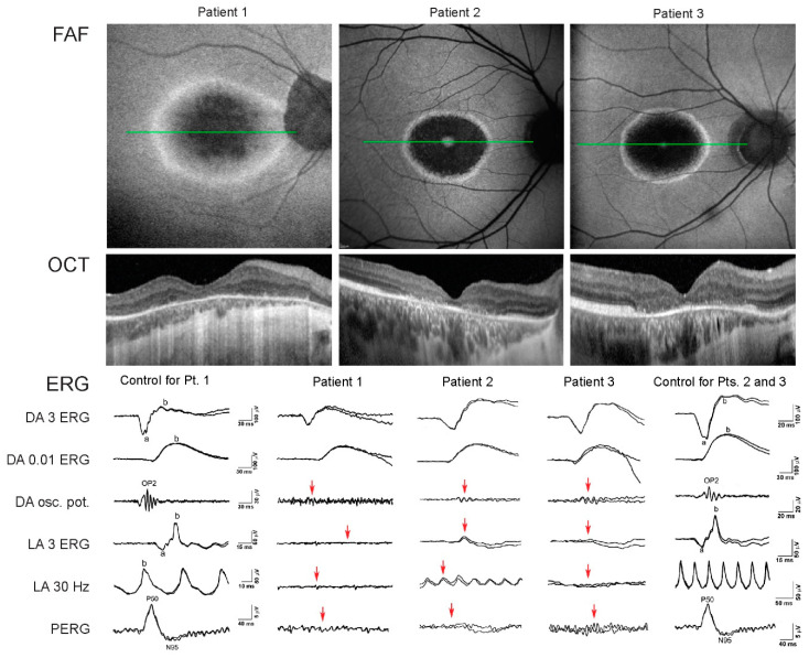
Mutations in are associated with rod-cone or cone/cone-rod dystrophy, the latter associated with mutations at the distal end. We describe the phenotype associated with a novel variant in the terminal codon of the c.3457T>A (Ter1153Lysext38), which results in a C-terminal extension. Three male patients from two families were recruited, aged 31, 35, and 38 years. Genetic testing was performed by whole exome sequencing. Filtered variants were analysed according to the population frequency, ClinVar database, the variant's putative impact, and predicted pathogenicity; and were classified according to the ACMG guidelines. Examination included visual acuity (Snellen), colour vision (Ishihara), visual field, fundus autofluorescence (FAF), optical coherence tomography (OCT), and electrophysiology. All patients were myopic, and had central scotoma and reduced colour vision. Visual acuities on better eyes were counting fingers, 0.3 and 0.05. Electrophysiology showed severely reduced cone-specific responses and macular dysfunction, while the rod-specific response was normal. FAF showed hyperautofluorescent ring centred at the fovea encompassing an area of photoreceptor loss approximately two optic discs in diameter (3462-6342 μm). Follow up after 2-11 years showed enlargement of the diameter (avg. 100 μm/year). The novel c.3457T>A (Ter1153Lysext38) mutation in the terminal codon is associated with cone dystrophy, which corresponds to the previously described phenotypes associated with mutations in the distal end of the . Minimal progression during follow-up years suggests a relatively stable disease after the initial loss of the central cones.
是与视杆细胞-视锥细胞或视锥细胞-视锥细胞营养不良相关的基因,后者与末端的突变有关。我们描述了一个新的末端密码子变异与相关的表型,c.3457T>A(Ter1153Lysext38),导致 C 端延伸。从两个家庭招募了 3 名男性患者,年龄分别为 31、35 和 38 岁。通过全外显子组测序进行基因检测。根据人群频率、ClinVar 数据库、变体的潜在影响和预测的致病性,对过滤后的变体进行分析;并根据 ACMG 指南进行分类。检查包括视力(Snellen)、色觉(Ishihara)、视野、眼底自发荧光(FAF)、光学相干断层扫描(OCT)和电生理学。所有患者均为近视,存在中心暗点和色觉减退。较好眼的视力分别为指数、0.3 和 0.05。电生理学显示锥细胞特异性反应严重降低,黄斑功能障碍,而杆细胞特异性反应正常。FAF 显示以黄斑为中心的高荧光环,包含一个直径约两个视盘(3462-6342μm)的光感受器丧失区。2-11 年的随访显示直径增大(平均每年 100μm)。末端密码子中的新型 c.3457T>A(Ter1153Lysext38)突变与锥细胞营养不良有关,这与以前描述的与末端突变相关的表型相对应。在随访期间的最小进展表明,在中央锥细胞丧失后,疾病相对稳定。