Department of Otorhinolaryngology-Head and Neck Surgery, School of Medicine University of Maryland, Baltimore, Maryland, United States.
Molecular Biology & Genetics Department, Liaquat University of Medical & Health Sciences, Jamshoro, Pakistan.
Invest Ophthalmol Vis Sci. 2019 Nov 1;60(14):4811-4819. doi: 10.1167/iovs.19-27263.
Cone rod dystrophy (CRD) is a group of inherited retinopathies characterized by the loss of cone and rod photoreceptor cells, which results in poor vision. This study aims to clinically and genetically characterize the segregating CRD phenotype in two large, consanguineous Pakistani families.
Funduscopy, optical coherence tomography (OCT), electroretinography (ERG), color vision, and visual acuity assessments were performed to evaluate the retinal structure and function of the affected individuals. Exome sequencing was performed to identify the genetic cause of CRD. Furthermore, the mutation's effect was evaluated using purified, bacterially expressed ADP-ribosylation factor-like protein 3 (ARL3) and mammalian cells.
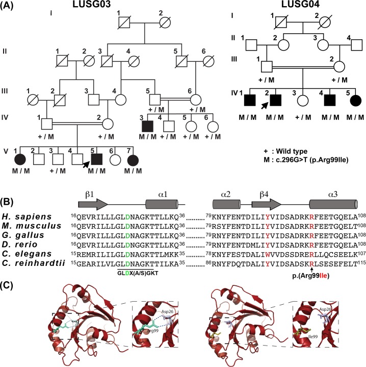
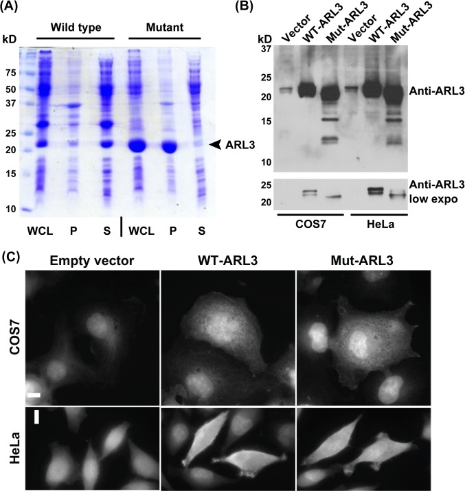
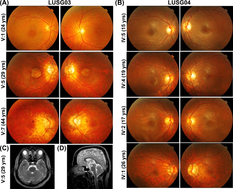
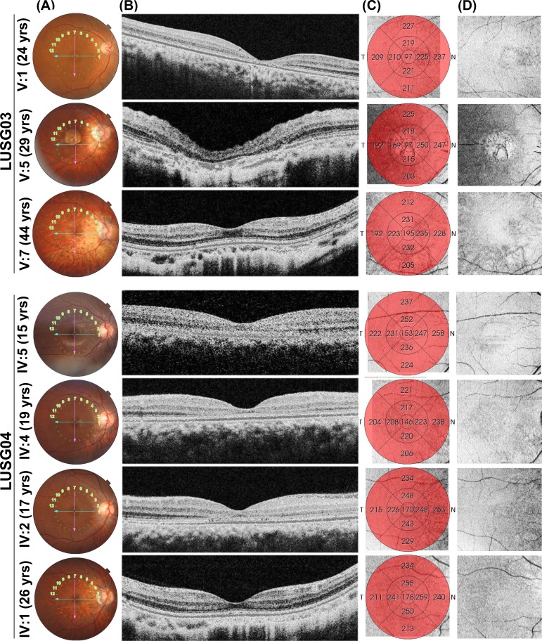
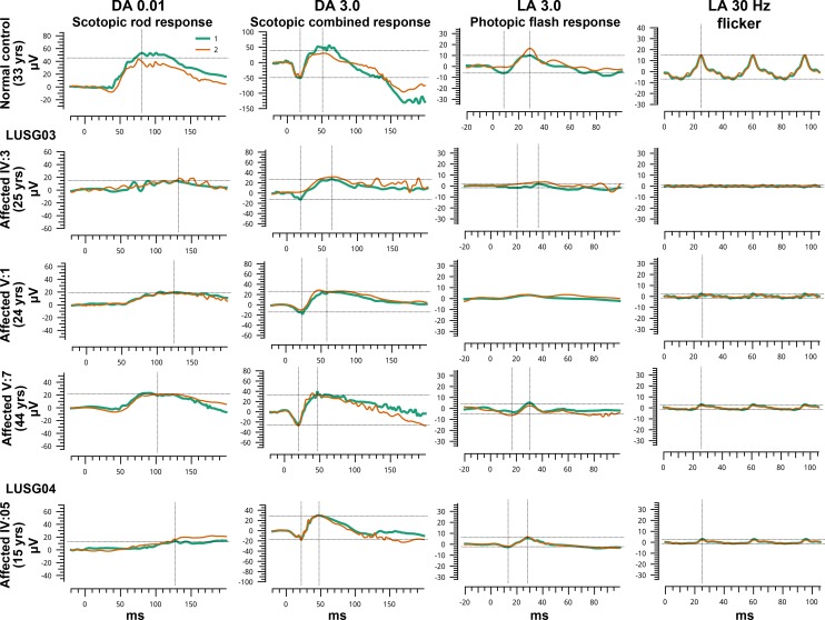
Fundus photography and OCT imaging demonstrated features that were consistent with CRD, including bull's eye macular lesions, macular atrophy, and central photoreceptor thinning. ERG analysis demonstrated moderate to severe reduction primarily of photopic responses in all affected individuals, and scotopic responses show reduction in two affected individuals. The exome sequencing revealed a novel homozygous variant (c.296G>T) in ARL3, which is predicted to substitute an evolutionarily conserved arginine with isoleucine within the encoded protein GTP-binding domain (R99I). The functional studies on the bacterial and heterologous mammalian cells revealed that the arginine at position 99 is essential for the stability of ARL3.
Our study uncovers an additional CRD gene and assigns the CRD phenotype to a variant of ARL3. The results imply that cargo transportation in photoreceptors as mediated by the ARL3 pathway is essential for cone and rod cell survival and vision in humans.
Cone rod dystrophy (CRD) 是一组遗传性视网膜病变,其特征是视锥细胞和视杆细胞的丧失,导致视力不佳。本研究旨在对两个大型近亲巴基斯坦家庭中分离出的 CRD 表型进行临床和遗传特征分析。
通过眼底检查、光学相干断层扫描 (OCT)、视网膜电图 (ERG)、色觉和视力评估来评估受影响个体的视网膜结构和功能。进行外显子组测序以确定 CRD 的遗传原因。此外,还使用纯化的、细菌表达的 ADP-ribosylation factor-like protein 3 (ARL3) 和哺乳动物细胞评估突变的影响。
眼底照相和 OCT 成像显示出与 CRD 一致的特征,包括牛眼黄斑病变、黄斑萎缩和中心感光细胞变薄。ERG 分析显示所有受影响个体的光反应均中度至重度降低,而两个受影响个体的暗反应降低。外显子组测序显示 ARL3 中存在一个新的纯合变异 (c.296G>T),该变异预计会在编码蛋白 GTP 结合域内将进化上保守的精氨酸替换为异亮氨酸 (R99I)。对细菌和异源哺乳动物细胞的功能研究表明,第 99 位的精氨酸对于 ARL3 的稳定性至关重要。
我们的研究揭示了另一个 CRD 基因,并将 CRD 表型归因于 ARL3 的变异。结果表明,由 ARL3 途径介导的货物运输对于视锥细胞和视杆细胞的存活以及人类的视力至关重要。