• 文献检索
  • 文档翻译
  • 深度研究
  • 学术资讯
  • Suppr Zotero 插件Zotero 插件
  • 邀请有礼
  • 套餐&价格
  • 历史记录
应用&插件
Suppr Zotero 插件Zotero 插件浏览器插件Mac 客户端Windows 客户端微信小程序
定价
高级版会员购买积分包购买API积分包
服务
文献检索文档翻译深度研究API 文档MCP 服务
关于我们
关于 Suppr公司介绍联系我们用户协议隐私条款
关注我们

Suppr 超能文献

核心技术专利:CN118964589B侵权必究
粤ICP备2023148730 号-1Suppr @ 2026

文献检索

告别复杂PubMed语法,用中文像聊天一样搜索,搜遍4000万医学文献。AI智能推荐,让科研检索更轻松。

立即免费搜索

文件翻译

保留排版,准确专业,支持PDF/Word/PPT等文件格式,支持 12+语言互译。

免费翻译文档

深度研究

AI帮你快速写综述,25分钟生成高质量综述,智能提取关键信息,辅助科研写作。

立即免费体验

关于影像学在新型冠状病毒肺炎检测、管理、组织病理学相关性及并发症中的作用的图文综述

A Pictorial Review of the Role of Imaging in the Detection, Management, Histopathological Correlations, and Complications of COVID-19 Pneumonia.

作者信息

Brogna Barbara, Bignardi Elio, Brogna Claudia, Volpe Mena, Lombardi Giulio, Rosa Alessandro, Gagliardi Giuliano, Capasso Pietro Fabio Maurizio, Gravino Enzo, Maio Francesca, Pane Francesco, Picariello Valentina, Buono Marcella, Colucci Lorenzo, Musto Lanfranco Aquilino

机构信息

Department of Radiology, San Giuseppe Moscati Hospital, Contrada Amoretta, 83100 Avellino, Italy.

Radiology Unit, Cotugno Hospital, Naples, Via Quagliariello 54, 80131 Naples, Italy.

出版信息

Diagnostics (Basel). 2021 Mar 4;11(3):437. doi: 10.3390/diagnostics11030437.

DOI:10.3390/diagnostics11030437
PMID:33806423
原文链接:https://pmc.ncbi.nlm.nih.gov/articles/PMC8000129/
Abstract

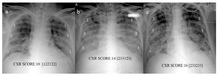
Imaging plays an important role in the detection of coronavirus (COVID-19) pneumonia in both managing the disease and evaluating the complications. Imaging with chest computed tomography (CT) can also have a potential predictive and prognostic role in COVID-19 patient outcomes. The aim of this pictorial review is to describe the role of imaging with chest X-ray (CXR), lung ultrasound (LUS), and CT in the diagnosis and management of COVID-19 pneumonia, the current indications, the scores proposed for each modality, the advantages/limitations of each modality and their role in detecting complications, and the histopathological correlations.

摘要

影像学在冠状病毒病(COVID-19)肺炎的检测中,对于疾病管理和并发症评估均发挥着重要作用。胸部计算机断层扫描(CT)成像在COVID-19患者的预后方面也可能具有潜在的预测和预后作用。本图片综述的目的是描述胸部X线(CXR)、肺部超声(LUS)和CT成像在COVID-19肺炎诊断和管理中的作用、当前适应症、针对每种检查方式提出的评分、每种检查方式的优缺点及其在检测并发症中的作用,以及组织病理学相关性。

https://cdn.ncbi.nlm.nih.gov/pmc/blobs/f226/8000129/77959b4c8037/diagnostics-11-00437-g013.jpg
https://cdn.ncbi.nlm.nih.gov/pmc/blobs/f226/8000129/58c78d90361c/diagnostics-11-00437-g001.jpg
https://cdn.ncbi.nlm.nih.gov/pmc/blobs/f226/8000129/5752f0516816/diagnostics-11-00437-g002.jpg
https://cdn.ncbi.nlm.nih.gov/pmc/blobs/f226/8000129/737fb450f1b2/diagnostics-11-00437-g003.jpg
https://cdn.ncbi.nlm.nih.gov/pmc/blobs/f226/8000129/5b4d639cd8df/diagnostics-11-00437-g004.jpg
https://cdn.ncbi.nlm.nih.gov/pmc/blobs/f226/8000129/42851712ed56/diagnostics-11-00437-g005.jpg
https://cdn.ncbi.nlm.nih.gov/pmc/blobs/f226/8000129/4ae457d9b5d2/diagnostics-11-00437-g006.jpg
https://cdn.ncbi.nlm.nih.gov/pmc/blobs/f226/8000129/1303c9bfccec/diagnostics-11-00437-g007.jpg
https://cdn.ncbi.nlm.nih.gov/pmc/blobs/f226/8000129/4e6ae816fe9f/diagnostics-11-00437-g008.jpg
https://cdn.ncbi.nlm.nih.gov/pmc/blobs/f226/8000129/b48e082498b2/diagnostics-11-00437-g009.jpg
https://cdn.ncbi.nlm.nih.gov/pmc/blobs/f226/8000129/665e058a5b7b/diagnostics-11-00437-g010.jpg
https://cdn.ncbi.nlm.nih.gov/pmc/blobs/f226/8000129/3de8bab60c1d/diagnostics-11-00437-g011.jpg
https://cdn.ncbi.nlm.nih.gov/pmc/blobs/f226/8000129/742a89a8a179/diagnostics-11-00437-g012.jpg
https://cdn.ncbi.nlm.nih.gov/pmc/blobs/f226/8000129/77959b4c8037/diagnostics-11-00437-g013.jpg

相似文献

1
A Pictorial Review of the Role of Imaging in the Detection, Management, Histopathological Correlations, and Complications of COVID-19 Pneumonia.关于影像学在新型冠状病毒肺炎检测、管理、组织病理学相关性及并发症中的作用的图文综述
Diagnostics (Basel). 2021 Mar 4;11(3):437. doi: 10.3390/diagnostics11030437.
2
Lung Ultrasound in COVID-19 Pneumonia: Correlations with Chest CT on Hospital admission.COVID-19 肺炎的肺部超声:与入院时胸部 CT 的相关性。
Respiration. 2020;99(7):617-624. doi: 10.1159/000509223. Epub 2020 Jun 22.
3
Lung Ultrasound in Patients With SARS-COV-2 Pneumonia: Correlations With Chest Computed Tomography, Respiratory Impairment, and Inflammatory Cascade.新型冠状病毒肺炎患者肺部超声:与胸部计算机断层扫描、呼吸障碍和炎症级联的相关性。
J Ultrasound Med. 2022 Jun;41(6):1465-1473. doi: 10.1002/jum.15831. Epub 2021 Sep 17.
4
The value of chest magnetic resonance imaging compared to chest radiographs with and without additional lung ultrasound in children with complicated pneumonia.胸部磁共振成像与常规 X 线胸片联合或不联合肺部超声在儿童复杂性肺炎中的应用价值。
PLoS One. 2020 Mar 19;15(3):e0230252. doi: 10.1371/journal.pone.0230252. eCollection 2020.
5
Low Sensitivity of Admission Lung US Compared to Chest CT for Diagnosis of Lung Involvement in a Cohort of 82 Patients with COVID-19 Pneumonia.在82例新冠肺炎患者队列中,与胸部CT相比,入院时肺部超声对诊断肺部受累的敏感性较低。
Medicina (Kaunas). 2021 Mar 4;57(3):236. doi: 10.3390/medicina57030236.
6
Lung Ultrasound vs. Chest X-Ray Study for the Radiographic Diagnosis of COVID-19 Pneumonia in a High-Prevalence Population.肺部超声与 X 线胸片对高流行地区 COVID-19 肺炎的放射学诊断比较。
J Emerg Med. 2021 May;60(5):615-625. doi: 10.1016/j.jemermed.2021.01.041. Epub 2021 Feb 4.
7
Comparative study of lung ultrasound and chest computed tomography scan in the assessment of severity of confirmed COVID-19 pneumonia.肺超声与胸部 CT 扫描在评估确诊 COVID-19 肺炎严重程度中的对比研究。
Intensive Care Med. 2020 Sep;46(9):1707-1713. doi: 10.1007/s00134-020-06186-0. Epub 2020 Jul 29.
8
Discrepancies in the clinical and radiological profiles of COVID-19: A case-based discussion and review of literature.新型冠状病毒肺炎临床与影像学特征的差异:基于病例的讨论及文献综述
World J Radiol. 2021 Apr 28;13(4):75-93. doi: 10.4329/wjr.v13.i4.75.
9
Prognostic Significance of Chest Imaging by LUS and CT in COVID-19 Inpatients: The ECOVID Multicenter Study.超声与 CT 肺部影像学对 COVID-19 住院患者预后评估的意义:ECCOVID 多中心研究。
Respiration. 2022;101(2):122-131. doi: 10.1159/000518516. Epub 2021 Aug 31.
10
Is Lung Ultrasound Imaging a Worthwhile Procedure for Severe Acute Respiratory Syndrome Coronavirus 2 Pneumonia Detection?肺部超声成像在严重急性呼吸综合征冠状病毒 2 型肺炎检测中是否有价值?
J Ultrasound Med. 2021 Jun;40(6):1113-1123. doi: 10.1002/jum.15487. Epub 2020 Sep 7.

引用本文的文献

1
Imaging of Acute Complications of Community-Acquired Pneumonia in the Paediatric Population-From Chest Radiography to MRI.儿童社区获得性肺炎急性并发症的影像学检查——从胸部X线摄影到磁共振成像
Children (Basel). 2024 Jan 18;11(1):122. doi: 10.3390/children11010122.
2
A Pictorial Essay Describing the CT Imaging Features of COVID-19 Cases throughout the Pandemic with a Special Focus on Lung Manifestations and Extrapulmonary Vascular Abdominal Complications.一篇图文并茂的文章,描述了新冠疫情期间新冠肺炎病例的CT成像特征,特别关注肺部表现和肺外血管腹部并发症。
Biomedicines. 2023 Jul 26;11(8):2113. doi: 10.3390/biomedicines11082113.
3

本文引用的文献

1
Pulmonary Vascular Manifestations of COVID-19 Pneumonia.新型冠状病毒肺炎的肺血管表现
Radiol Cardiothorac Imaging. 2020 Jun 18;2(3):e200277. doi: 10.1148/ryct.2020200277. eCollection 2020 Jun.
2
Lethal COVID-19: Radiologic-Pathologic Correlation of the Lungs.致命性新冠肺炎:肺部的放射学与病理学相关性
Radiol Cardiothorac Imaging. 2020 Nov 19;2(6):e200406. doi: 10.1148/ryct.2020200406. eCollection 2020 Dec.
3
Prognostic Value and Reproducibility of AI-assisted Analysis of Lung Involvement in COVID-19 on Low-Dose Submillisievert Chest CT: Sample Size Implications for Clinical Trials.
Earliest histopathological changes in COVID-19 pneumonia with comprehensive gene expression analyses: A case series study.
伴有综合基因表达分析的 COVID-19 肺炎的最早组织病理学改变:一项病例系列研究。
Histol Histopathol. 2023 Jun;38(6):623-636. doi: 10.14670/HH-18-557. Epub 2022 Dec 1.
4
Artificial Intelligence-Based Detection of Pneumonia in Chest Radiographs.基于人工智能的胸部X光片中肺炎检测
Diagnostics (Basel). 2022 Jun 14;12(6):1465. doi: 10.3390/diagnostics12061465.
5
Imaging-based indices combining disease severity and time from disease onset to predict COVID-19 mortality: A cohort study.基于影像学的疾病严重程度和从发病到预测 COVID-19 死亡率的时间的综合指数:一项队列研究。
PLoS One. 2022 Jun 16;17(6):e0270111. doi: 10.1371/journal.pone.0270111. eCollection 2022.
6
Spontaneous pneumothorax, pneumomediastinum and subcutaneous emphysema in non-ventilated COVID-19 patients.非通气型新冠病毒肺炎患者的自发性气胸、纵隔气肿和皮下气肿
Future Sci OA. 2021 Nov 18;8(2):FSO771. doi: 10.2144/fsoa-2021-0090. eCollection 2022 Feb.
7
Differences in Dynamics of Lung Computed Tomography Patterns between Survivors and Deceased Adult Patients with COVID-19.新冠病毒肺炎成年幸存者与死亡患者肺部计算机断层扫描模式动态变化的差异
Diagnostics (Basel). 2021 Oct 19;11(10):1937. doi: 10.3390/diagnostics11101937.
8
Perfusion Patterns of Peripheral Organizing Pneumonia (POP) Using Contrast-Enhanced Ultrasound (CEUS) and Their Correlation with Immunohistochemically Detected Vascularization Patterns.使用超声造影(CEUS)评估外周型机化性肺炎(POP)的灌注模式及其与免疫组化检测的血管生成模式的相关性
Diagnostics (Basel). 2021 Sep 2;11(9):1601. doi: 10.3390/diagnostics11091601.
9
COVID-19 Pneumonia: The Great Ultrasonography Mimicker.新型冠状病毒肺炎:超声检查的强大模仿者
Front Med (Lausanne). 2021 Aug 25;8:709402. doi: 10.3389/fmed.2021.709402. eCollection 2021.
10
Lung Ultrasound in the Diagnosis of COVID-19 Pneumonia: Not Always and Not Only What Is COVID-19 "Glitters".肺部超声在COVID-19肺炎诊断中的应用:并非总是且不仅仅是COVID-19的“闪光点”。
Front Med (Lausanne). 2021 Jul 19;8:707602. doi: 10.3389/fmed.2021.707602. eCollection 2021.
人工智能辅助分析低剂量亚毫西弗胸部CT上COVID-19肺部受累情况的预后价值及可重复性:对临床试验样本量的影响
Radiol Cardiothorac Imaging. 2020 Oct 22;2(5):e200441. doi: 10.1148/ryct.2020200441. eCollection 2020 Oct.
4
Chest CT Severity Score: An Imaging Tool for Assessing Severe COVID-19.胸部CT严重程度评分:一种评估重症COVID-19的影像学工具。
Radiol Cardiothorac Imaging. 2020 Mar 30;2(2):e200047. doi: 10.1148/ryct.2020200047. eCollection 2020 Apr.
5
Prognostic value of bedside lung ultrasound score in patients with COVID-19.COVID-19 患者床边肺部超声评分的预后价值。
Crit Care. 2020 Dec 22;24(1):700. doi: 10.1186/s13054-020-03416-1.
6
COVID-19 and Spontaneous Pneumomediastinum: A case series.新型冠状病毒肺炎与自发性纵隔气肿:病例系列
Heart Lung. 2021 Mar-Apr;50(2):202-205. doi: 10.1016/j.hrtlng.2020.12.002. Epub 2020 Dec 4.
7
Predictive value of chest CT scoring in COVID-19 patients in Wuhan, China: A retrospective cohort study.中国武汉 COVID-19 患者的胸部 CT 评分的预测价值:一项回顾性队列研究。
Respir Med. 2021 Jan;176:106271. doi: 10.1016/j.rmed.2020.106271. Epub 2020 Nov 28.
8
Interstitial lung opacities in patients with severe COVID-19 pneumonia by bedside high-resolution ultrasound in association to CO2 retention.严重 COVID-19 肺炎患者的间质肺混浊通过床边高分辨率超声与 CO2 潴留相关。
Clin Hemorheol Microcirc. 2021;77(4):355-365. doi: 10.3233/CH-200925.
9
Which role for chest x-ray score in predicting the outcome in COVID-19 pneumonia?胸部X光评分在预测COVID-19肺炎预后方面起何种作用?
Eur Radiol. 2021 Jun;31(6):4016-4022. doi: 10.1007/s00330-020-07504-2. Epub 2020 Dec 2.
10
Two cases of spontaneous pneumomediastinum with pneumothorax in patients with COVID-19 associated pneumonia.两例新型冠状病毒肺炎相关肺炎患者并发自发性纵隔气肿伴气胸
Respir Med Case Rep. 2020;31:101308. doi: 10.1016/j.rmcr.2020.101308. Epub 2020 Nov 26.