• 文献检索
  • 文档翻译
  • 深度研究
  • 学术资讯
  • Suppr Zotero 插件Zotero 插件
  • 邀请有礼
  • 套餐&价格
  • 历史记录
应用&插件
Suppr Zotero 插件Zotero 插件浏览器插件Mac 客户端Windows 客户端微信小程序
定价
高级版会员购买积分包购买API积分包
服务
文献检索文档翻译深度研究API 文档MCP 服务
关于我们
关于 Suppr公司介绍联系我们用户协议隐私条款
关注我们

Suppr 超能文献

核心技术专利:CN118964589B侵权必究
粤ICP备2023148730 号-1Suppr @ 2026

文献检索

告别复杂PubMed语法,用中文像聊天一样搜索,搜遍4000万医学文献。AI智能推荐,让科研检索更轻松。

立即免费搜索

文件翻译

保留排版,准确专业,支持PDF/Word/PPT等文件格式,支持 12+语言互译。

免费翻译文档

深度研究

AI帮你快速写综述,25分钟生成高质量综述,智能提取关键信息,辅助科研写作。

立即免费体验

新冠病毒肺炎成年幸存者与死亡患者肺部计算机断层扫描模式动态变化的差异

Differences in Dynamics of Lung Computed Tomography Patterns between Survivors and Deceased Adult Patients with COVID-19.

作者信息

Akopyan Gevorg B, Berdalin Alexander B, Gubskiy Ilya L, Lelyuk Vladimir G

机构信息

Radiology and Clinical Physiology Research Center, Federal State Budgetary Institution Federal Center of Brain Research and Neurotechnologies of the Federal Medical Biological Agency, 117513 Moscow, Russia.

出版信息

Diagnostics (Basel). 2021 Oct 19;11(10):1937. doi: 10.3390/diagnostics11101937.

DOI:10.3390/diagnostics11101937
PMID:34679635
原文链接:https://pmc.ncbi.nlm.nih.gov/articles/PMC8534345/
Abstract

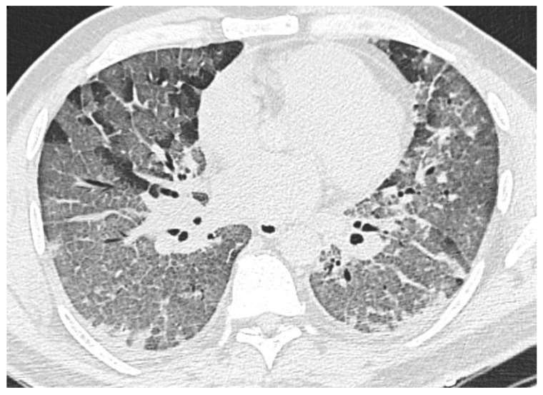
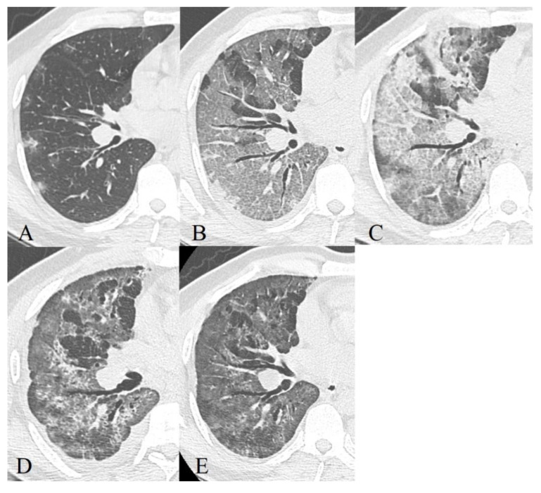
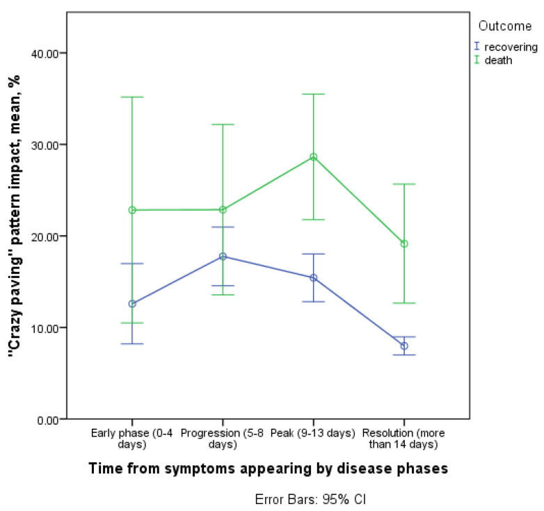
This study's aim was to investigate CT (computed tomography) pattern dynamics differences within surviving and deceased adult patients with COVID-19, revealing new prognostic factors and reproducing already known data with our patients' cohort: 635 hospitalized patients (55.3% of them were men, 44.7%-women), of which 87.3% had a positive result of RT-PCR (reverse transcription-polymerase chain reaction) at admission. The number of deaths was 53 people (69.8% of them were men and 30.2% were women). In total, more than 1500 CT examinations were performed on patients, using a GE Optima CT 660 computed tomography (General Electric Healthcare, Chicago, IL, USA). The study was performed at hospital admission, the frequency of repetitive scans further varied based on clinical need. The interpretation of the imaging data was carried out by 11 radiologists with filling in individual registration cards that take into account the scale of the lesion, the location, contours, and shape of the foci, the dominating types of changes, as well as the presence of additional findings and the dynamics of the process-a total of 45 parameters. Statistical analysis was performed using the software packages SPSS Statistics version 23.0 (IBM, Armonk, NY, USA) and R software version 3.3.2. For comparisons in pattern dynamics across hospitalization we used repeated measures general linear model with outcome and disease phase as factors. The crazy paving pattern, which is more common and has a greater contribution to the overall CT picture in different phases of the disease in deceased patients, has isolated prognostic significance and is probably a reflection of faster dynamics of the process with a long phase of progression of pulmonary parenchyma damage with an identical trend of changes in the scale of the lesion (as recovered) in this group of patients. Already known data on typical pulmonological CT manifestations of infection, frequency of occurrence, and the prognostic significance of the scale of the lesion were reproduced, new differences in the dynamics of the process between recovered and deceased adult patients were also found that may have prognostic significance and can be reflected in clinical practice.

摘要

本研究旨在调查新型冠状病毒肺炎(COVID-19)成年存活患者与死亡患者的CT(计算机断层扫描)影像动态差异,揭示新的预后因素,并通过我们的患者队列重现已知数据:635例住院患者(其中55.3%为男性,44.7%为女性),其中87.3%在入院时逆转录聚合酶链反应(RT-PCR)结果呈阳性。死亡人数为53人(其中69.8%为男性,30.2%为女性)。总共对患者进行了1500多次CT检查,使用的是GE Optima CT 660计算机断层扫描仪(美国伊利诺伊州芝加哥市通用电气医疗集团)。该研究在患者入院时进行,重复扫描的频率根据临床需要进一步变化。11名放射科医生对影像数据进行解读,并填写个人登记卡,登记卡考虑了病变范围、病灶位置、轮廓和形状、主要变化类型,以及是否存在其他发现和病情动态变化——总共45个参数。使用SPSS Statistics 23.0软件包(美国纽约州阿蒙克市IBM公司)和R软件3.3.2版本进行统计分析。为了比较住院期间的影像动态变化,我们使用了以结果和疾病阶段为因素的重复测量一般线性模型。碎石路征在死亡患者疾病的不同阶段更常见,对整体CT影像的贡献更大,具有独立的预后意义,可能反映了该组患者病程进展更快,肺实质损伤进展期较长,病变范围(恢复时)变化趋势相同。重现了关于感染典型肺部CT表现、发生率以及病变范围预后意义的已知数据,还发现了康复成年患者与死亡成年患者病程动态变化的新差异,这些差异可能具有预后意义,并可在临床实践中得到体现。

https://cdn.ncbi.nlm.nih.gov/pmc/blobs/fcec/8534345/5cc81a3621d5/diagnostics-11-01937-g015.jpg
https://cdn.ncbi.nlm.nih.gov/pmc/blobs/fcec/8534345/9befe4902d87/diagnostics-11-01937-g001.jpg
https://cdn.ncbi.nlm.nih.gov/pmc/blobs/fcec/8534345/80e1f6e836c2/diagnostics-11-01937-g002.jpg
https://cdn.ncbi.nlm.nih.gov/pmc/blobs/fcec/8534345/2525397d1f14/diagnostics-11-01937-g003.jpg
https://cdn.ncbi.nlm.nih.gov/pmc/blobs/fcec/8534345/177512113fbc/diagnostics-11-01937-g004.jpg
https://cdn.ncbi.nlm.nih.gov/pmc/blobs/fcec/8534345/050735a4ee1b/diagnostics-11-01937-g005.jpg
https://cdn.ncbi.nlm.nih.gov/pmc/blobs/fcec/8534345/c21535e4d74f/diagnostics-11-01937-g006.jpg
https://cdn.ncbi.nlm.nih.gov/pmc/blobs/fcec/8534345/0662e2124e40/diagnostics-11-01937-g007.jpg
https://cdn.ncbi.nlm.nih.gov/pmc/blobs/fcec/8534345/79b2bc00c07f/diagnostics-11-01937-g008.jpg
https://cdn.ncbi.nlm.nih.gov/pmc/blobs/fcec/8534345/5f9e30622f0c/diagnostics-11-01937-g009.jpg
https://cdn.ncbi.nlm.nih.gov/pmc/blobs/fcec/8534345/ba9f97f3fc5b/diagnostics-11-01937-g010.jpg
https://cdn.ncbi.nlm.nih.gov/pmc/blobs/fcec/8534345/7a54f305d415/diagnostics-11-01937-g011.jpg
https://cdn.ncbi.nlm.nih.gov/pmc/blobs/fcec/8534345/96513a9d66dd/diagnostics-11-01937-g012.jpg
https://cdn.ncbi.nlm.nih.gov/pmc/blobs/fcec/8534345/b70705c10faa/diagnostics-11-01937-g013.jpg
https://cdn.ncbi.nlm.nih.gov/pmc/blobs/fcec/8534345/9afb996c8b83/diagnostics-11-01937-g014.jpg
https://cdn.ncbi.nlm.nih.gov/pmc/blobs/fcec/8534345/5cc81a3621d5/diagnostics-11-01937-g015.jpg

相似文献

1
Differences in Dynamics of Lung Computed Tomography Patterns between Survivors and Deceased Adult Patients with COVID-19.新冠病毒肺炎成年幸存者与死亡患者肺部计算机断层扫描模式动态变化的差异
Diagnostics (Basel). 2021 Oct 19;11(10):1937. doi: 10.3390/diagnostics11101937.
2
[Spatial and temporal distribution and predictive value of chest CT scoring in patients with COVID-19].新型冠状病毒肺炎患者胸部CT评分的时空分布及预测价值
Zhonghua Jie He He Hu Xi Za Zhi. 2021 Mar 12;44(3):230-236. doi: 10.3760/cma.j.cn112147-20200522-00626.
3
Frequency of typical and atypical computed tomography findings of COVID-19 and their effect on hospitalization.新型冠状病毒肺炎(COVID-19)典型与非典型计算机断层扫描表现的频率及其对住院治疗的影响。
North Clin Istanb. 2021 Dec 29;8(6):554-561. doi: 10.14744/nci.2021.24865. eCollection 2021.
4
CT Features of COVID-19 Pneumonia Differ Depending on the Severity and Duration of Disease.新冠肺炎肺炎的 CT 特征取决于疾病的严重程度和持续时间。
Rofo. 2021 Jun;193(6):672-682. doi: 10.1055/a-1293-9163. Epub 2020 Dec 17.
5
CT imaging features of COVID-19 pneumonia: initial experience from Turkey.土耳其的 COVID-19 肺炎 CT 影像学特征:初步经验。
Diagn Interv Radiol. 2020 Jul;26(4):308-314. doi: 10.5152/dir.2020.20307.
6
Temporal relationship between serial RT-PCR results and serial chest CT imaging, and serial CT changes in coronavirus 2019 (COVID-19) pneumonia: a descriptive study of 155 cases in China.新型冠状病毒(COVID-19)肺炎的实时逆转录聚合酶链反应(RT-PCR)结果与系列胸部 CT 影像学及系列 CT 改变的时间关系:中国 155 例的描述性研究。
Eur Radiol. 2021 Mar;31(3):1175-1184. doi: 10.1007/s00330-020-07268-9. Epub 2020 Sep 15.
7
Pregnancy and Perinatal Outcomes of Women With Coronavirus Disease (COVID-19) Pneumonia: A Preliminary Analysis.妊娠期和围产期新型冠状病毒肺炎患者的结局:初步分析。
AJR Am J Roentgenol. 2020 Jul;215(1):127-132. doi: 10.2214/AJR.20.23072. Epub 2020 Mar 18.
8
Evaluation of initial chest computed tomography (CT) findings of COVID-19 pneumonia in 117 deceased patients: a retrospective study.117例死亡的新型冠状病毒肺炎患者初始胸部计算机断层扫描(CT)表现的评估:一项回顾性研究
Turk J Med Sci. 2021 Jun 28;51(3):929-938. doi: 10.3906/sag-2009-183.
9
Early Clinical and CT Manifestations of Coronavirus Disease 2019 (COVID-19) Pneumonia.新型冠状病毒肺炎(COVID-19)肺炎的早期临床和 CT 表现。
AJR Am J Roentgenol. 2020 Aug;215(2):338-343. doi: 10.2214/AJR.20.22961. Epub 2020 Mar 17.
10
Temporal Changes of CT Findings in 90 Patients with COVID-19 Pneumonia: A Longitudinal Study.90 例 COVID-19 肺炎患者 CT 表现的时间变化:一项纵向研究。
Radiology. 2020 Aug;296(2):E55-E64. doi: 10.1148/radiol.2020200843. Epub 2020 Mar 19.

本文引用的文献

1
COVID-19 Pneumonia in Vaccinated Population: A Six Clinical and Radiological Case Series.接种人群中的 COVID-19 肺炎:六例临床和放射学病例系列。
Medicina (Kaunas). 2021 Aug 27;57(9):891. doi: 10.3390/medicina57090891.
2
Risk Factors of In-Hospital Mortality in Non-Specialized Tertiary Center Repurposed for Medical Care to COVID-19 Patients in Russia.俄罗斯将非专科三级医疗中心改造用于治疗新冠患者时院内死亡的危险因素
Diagnostics (Basel). 2021 Sep 15;11(9):1687. doi: 10.3390/diagnostics11091687.
3
Reshaping of Italian Echocardiographic Laboratories Activities during the Second Wave of COVID-19 Pandemic and Expectations for the Post-Pandemic Era.
意大利超声心动图实验室在新冠疫情第二波期间的活动重塑及对疫情后时代的期望
J Clin Med. 2021 Aug 5;10(16):3466. doi: 10.3390/jcm10163466.
4
Evolution of CT Findings and Lung Residue in Patients with COVID-19 Pneumonia: Quantitative Analysis of the Disease with a Computer Automatic Tool.新型冠状病毒肺炎患者CT表现及肺残留灶的演变:使用计算机自动工具对疾病进行定量分析
J Pers Med. 2021 Jul 6;11(7):641. doi: 10.3390/jpm11070641.
5
A Pictorial Review of the Role of Imaging in the Detection, Management, Histopathological Correlations, and Complications of COVID-19 Pneumonia.关于影像学在新型冠状病毒肺炎检测、管理、组织病理学相关性及并发症中的作用的图文综述
Diagnostics (Basel). 2021 Mar 4;11(3):437. doi: 10.3390/diagnostics11030437.
6
Lethal COVID-19: Radiologic-Pathologic Correlation of the Lungs.致命性新冠肺炎:肺部的放射学与病理学相关性
Radiol Cardiothorac Imaging. 2020 Nov 19;2(6):e200406. doi: 10.1148/ryct.2020200406. eCollection 2020 Dec.
7
Thin-Section Chest CT Imaging of COVID-19 Pneumonia: A Comparison Between Patients with Mild and Severe Disease.新型冠状病毒肺炎的胸部薄层CT成像:轻型与重型患者的比较
Radiol Cardiothorac Imaging. 2020 Apr 23;2(2):e200126. doi: 10.1148/ryct.2020200126. eCollection 2020 Apr.
8
Chest CT Findings in Cases from the Cruise Ship with Coronavirus Disease (COVID-19).邮轮上新型冠状病毒肺炎(COVID-19)病例的胸部CT表现
Radiol Cardiothorac Imaging. 2020 Mar 17;2(2):e200110. doi: 10.1148/ryct.2020200110. eCollection 2020 Apr.
9
Chest CT Severity Score: An Imaging Tool for Assessing Severe COVID-19.胸部CT严重程度评分:一种评估重症COVID-19的影像学工具。
Radiol Cardiothorac Imaging. 2020 Mar 30;2(2):e200047. doi: 10.1148/ryct.2020200047. eCollection 2020 Apr.
10
Imaging Profile of the COVID-19 Infection: Radiologic Findings and Literature Review.新型冠状病毒肺炎感染的影像学表现:放射学发现与文献综述
Radiol Cardiothorac Imaging. 2020 Feb 13;2(1):e200034. doi: 10.1148/ryct.2020200034. eCollection 2020 Feb.