Becker Judith, Decker Josua A, Römmele Christoph, Kahn Maria, Messmann Helmut, Wehler Markus, Schwarz Florian, Kroencke Thomas, Scheurig-Muenkler Christian
Department of Diagnostic and Interventional Radiology and Neuroradiology, University Hospital Augsburg, Stenglinstraße 2, 86156 Augsburg, Germany.
Department of Gastroenterology, University Hospital Augsburg, Stenglinstraße 2, 86156 Augsburg, Germany.
Diagnostics (Basel). 2022 Jun 14;12(6):1465. doi: 10.3390/diagnostics12061465.
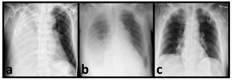
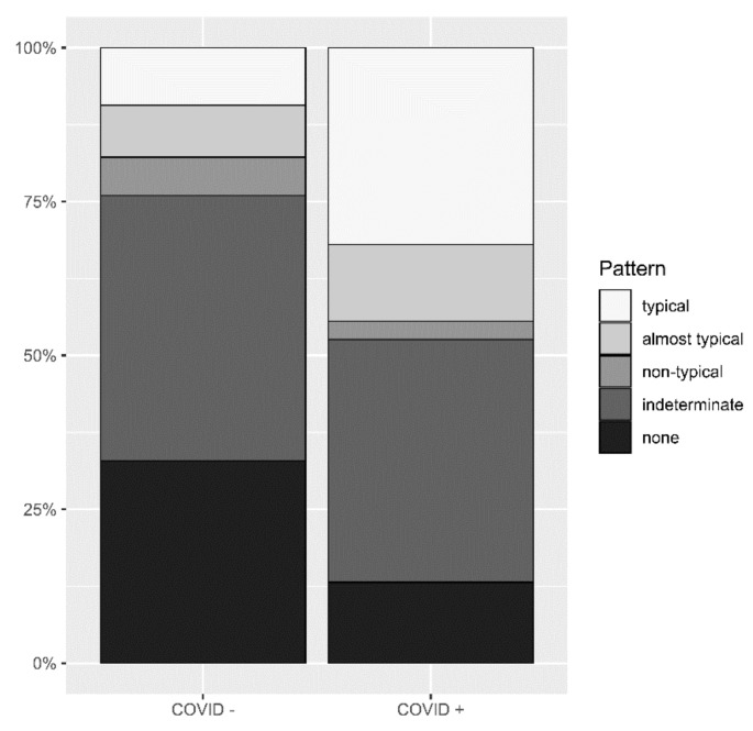
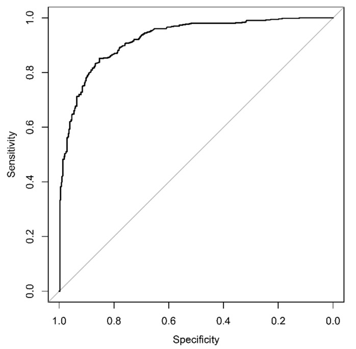
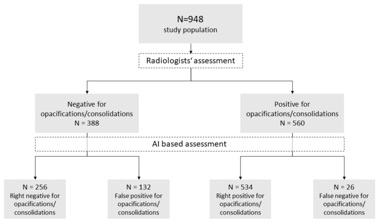
Artificial intelligence is gaining increasing relevance in the field of radiology. This study retrospectively evaluates how a commercially available deep learning algorithm can detect pneumonia in chest radiographs (CR) in emergency departments. The chest radiographs of 948 patients with dyspnea between 3 February and 8 May 2020, as well as 15 October and 15 December 2020, were used. A deep learning algorithm was used to identify opacifications associated with pneumonia, and the performance was evaluated by using ROC analysis, sensitivity, specificity, PPV and NPV. Two radiologists assessed all enrolled images for pulmonal infection patterns as the reference standard. If consolidations or opacifications were present, the radiologists classified the pulmonal findings regarding a possible COVID-19 infection because of the ongoing pandemic. The AUROC value of the deep learning algorithm reached 0.923 when detecting pneumonia in chest radiographs with a sensitivity of 95.4%, specificity of 66.0%, PPV of 80.2% and NPV of 90.8%. The detection of COVID-19 pneumonia in CR by radiologists was achieved with a sensitivity of 50.6% and a specificity of 73%. The deep learning algorithm proved to be an excellent tool for detecting pneumonia in chest radiographs. Thus, the assessment of suspicious chest radiographs can be purposefully supported, shortening the turnaround time for reporting relevant findings and aiding early triage.
人工智能在放射学领域正变得越来越重要。本研究回顾性评估了一种商用深度学习算法在急诊科胸部X光片(CR)中检测肺炎的能力。研究使用了2020年2月3日至5月8日以及10月15日至12月15日期间948例呼吸困难患者的胸部X光片。使用深度学习算法识别与肺炎相关的肺部混浊,并通过ROC分析、灵敏度、特异性、阳性预测值和阴性预测值评估其性能。两名放射科医生将所有纳入的图像评估为肺部感染模式作为参考标准。由于疫情仍在持续,如果存在实变或混浊,放射科医生会对肺部检查结果进行分类,以判断是否可能感染了COVID-19。在胸部X光片中检测肺炎时,深度学习算法的AUROC值达到0.923,灵敏度为95.4%,特异性为66.0%,阳性预测值为80.2%,阴性预测值为90.8%。放射科医生在CR中检测COVID-19肺炎的灵敏度为50.6%,特异性为73%。深度学习算法被证明是检测胸部X光片中肺炎的优秀工具。因此,可以有针对性地支持对可疑胸部X光片的评估,缩短报告相关结果的周转时间,并有助于早期分诊。