Henkel Maurice, Weikert Thomas, Marston Katharina, Schwab Nathalie, Sommer Gregor, Haslbauer Jasmin, Franzeck Fabian, Anastasopoulos Constantin, Stieltjes Bram, Michel Anne, Bremerich Jens, Menter Thomas, Mertz Kirsten D, Tzankov Alexandar, Sauter Alexander W
Department of Radiology (M.H., T.W., G.S., C.A., B.S., J.B., A.W.S.), Department of Research & Analytic Services (M.H., T.W., F.F., B.S.), and Division of Histopathology and Autopsy, Institute of Pathology (K.M., J.H., T.M., A.T.), University Hospital Basel, University of Basel, Petersgraben 4, 4031 Basel, Switzerland; and Department of Pathology, Cantonal Hospital Baselland, Liestal, Switzerland (M.H., N.S., A.M., K.D.M.).
Radiol Cardiothorac Imaging. 2020 Nov 19;2(6):e200406. doi: 10.1148/ryct.2020200406. eCollection 2020 Dec.
The purpose of this retrospective study was to correlate CT patterns of fatal cases of coronavirus disease 2019 (COVID-19) with postmortem pathology observations.
The study included 70 lung lobes of 14 patients who died of reverse-transcription polymerase chain reaction-confirmed COVID-19. All patients underwent antemortem CT and autopsy between March 9 and April 30, 2020. Board-certified radiologists and pathologists performed lobewise correlations of pulmonary observations. In a consensus reading, 267 radiologic and 257 histopathologic observations of the lungs were recorded and systematically graded according to severity. These observations were matched and evaluated.
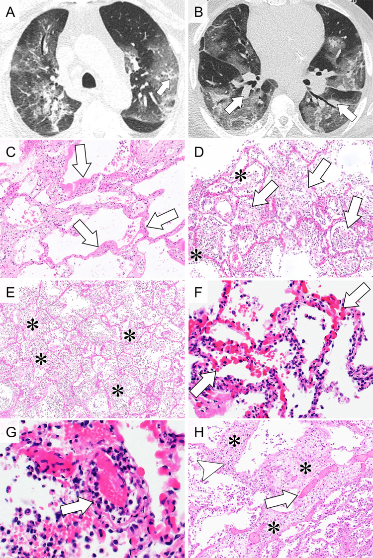
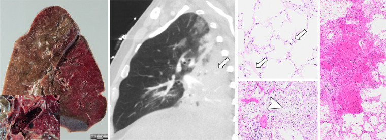
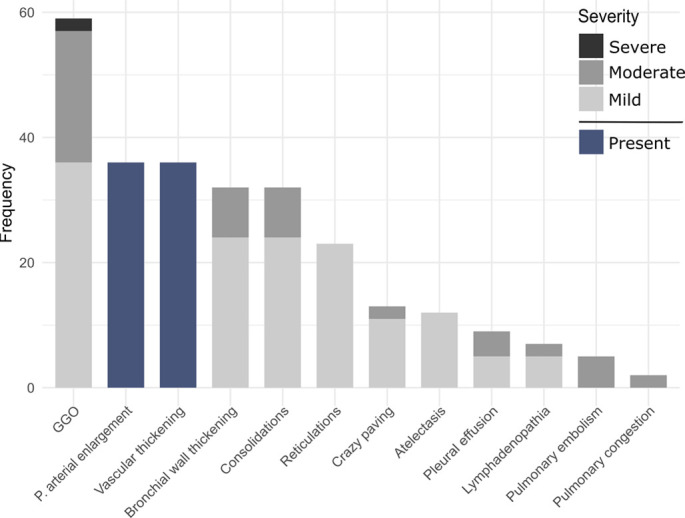
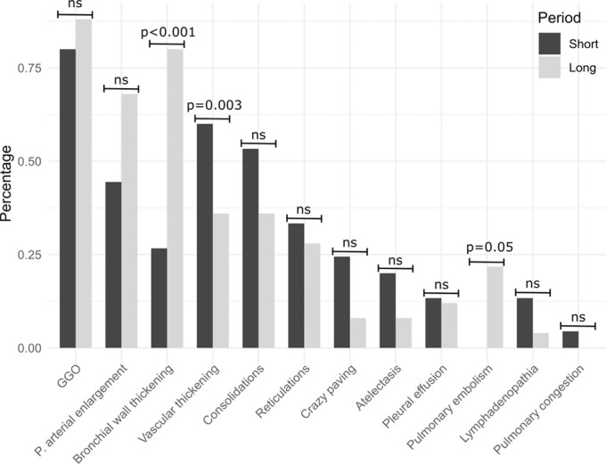
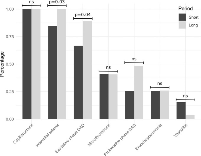
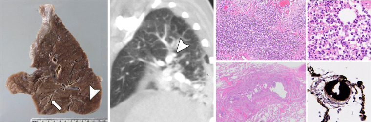
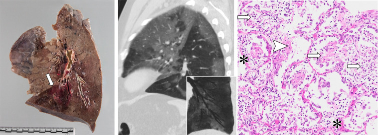
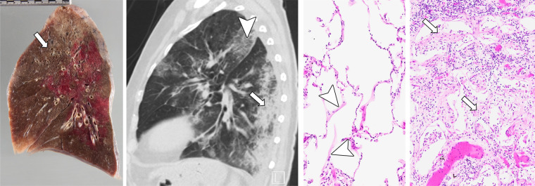
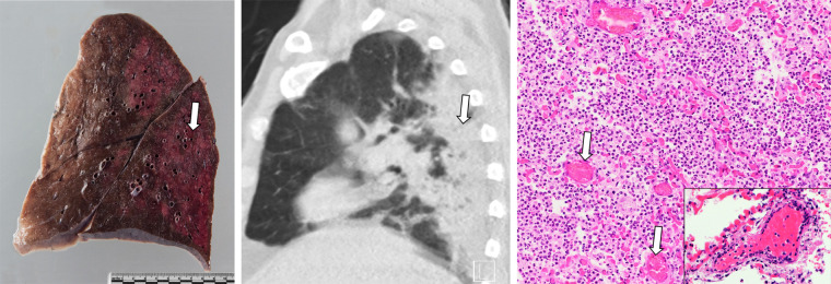
Predominant CT observations were ground-glass opacities (GGO) (59/70 lobes examined) and areas of consolidation (33/70). The histopathologic observations were consistent with diffuse alveolar damage (70/70) and capillary dilatation and congestion (70/70), often accompanied by microthrombi (27/70), superimposed acute bronchopneumonia (17/70), and leukocytoclastic vasculitis (7/70). Four patients had pulmonary emboli. Bronchial wall thickening at CT histologically corresponded with acute bronchopneumonia. GGOs and consolidations corresponded with mixed histopathologic observations, including capillary dilatation and congestion, interstitial edema, diffuse alveolar damage, and microthrombosis. Vascular alterations were prominent observations at both CT and histopathology.
A significant proportion of GGO correlated with the pathologic processes of diffuse alveolar damage, capillary dilatation and congestion, and microthrombosis. Our results confirm the presence and underline the importance of vascular alterations as key pathophysiologic drivers in lethal COVID-19.© RSNA, 2020.
本回顾性研究旨在将2019冠状病毒病(COVID-19)死亡病例的CT表现与尸检病理观察结果进行关联。
本研究纳入了14例经逆转录聚合酶链反应确诊死于COVID-19患者的70个肺叶。所有患者于2020年3月9日至4月30日期间接受了生前CT检查和尸检。经过认证的放射科医生和病理科医生对肺部观察结果进行了分叶关联。在一次共识读片中,记录了267项肺部放射学观察结果和257项组织病理学观察结果,并根据严重程度进行了系统分级。对这些观察结果进行了匹配和评估。
主要的CT表现为磨玻璃影(GGO)(70个检查肺叶中的59个)和实变区域(70个中的33个)。组织病理学观察结果与弥漫性肺泡损伤(70/70)、毛细血管扩张和充血(70/70)一致,常伴有微血栓形成(27/70)、叠加的急性支气管肺炎(17/70)和白细胞破碎性血管炎(7/70)。4例患者有肺栓塞。CT表现的支气管壁增厚在组织学上与急性支气管肺炎相符。GGO和实变与混合的组织病理学观察结果相符,包括毛细血管扩张和充血、间质水肿、弥漫性肺泡损伤和微血栓形成。血管改变在CT和组织病理学检查中均为突出表现。
相当一部分GGO与弥漫性肺泡损伤、毛细血管扩张和充血以及微血栓形成的病理过程相关。我们的结果证实了血管改变的存在,并强调了其作为致死性COVID-19关键病理生理驱动因素的重要性。© RSNA,2020。