• 文献检索
  • 文档翻译
  • 深度研究
  • 学术资讯
  • Suppr Zotero 插件Zotero 插件
  • 邀请有礼
  • 套餐&价格
  • 历史记录
应用&插件
Suppr Zotero 插件Zotero 插件浏览器插件Mac 客户端Windows 客户端微信小程序
定价
高级版会员购买积分包购买API积分包
服务
文献检索文档翻译深度研究API 文档MCP 服务
关于我们
关于 Suppr公司介绍联系我们用户协议隐私条款
关注我们

Suppr 超能文献

核心技术专利:CN118964589B侵权必究
粤ICP备2023148730 号-1Suppr @ 2026

文献检索

告别复杂PubMed语法,用中文像聊天一样搜索,搜遍4000万医学文献。AI智能推荐,让科研检索更轻松。

立即免费搜索

文件翻译

保留排版,准确专业,支持PDF/Word/PPT等文件格式,支持 12+语言互译。

免费翻译文档

深度研究

AI帮你快速写综述,25分钟生成高质量综述,智能提取关键信息,辅助科研写作。

立即免费体验

骨 肉 瘤 肺 转 移 的 放 射 学 评 估 : 现 状 和 最 新 进 展 。

Radiologic Assessment of Osteosarcoma Lung Metastases: State of the Art and Recent Advances.

机构信息

Diagnostic and Interventional Radiology Unit, IRCCS Istituto Ortopedico Rizzoli, 40136 Bologna, Italy.

出版信息

Cells. 2021 Mar 4;10(3):553. doi: 10.3390/cells10030553.

DOI:10.3390/cells10030553
PMID:33806513
原文链接:https://pmc.ncbi.nlm.nih.gov/articles/PMC7999261/
Abstract

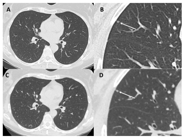
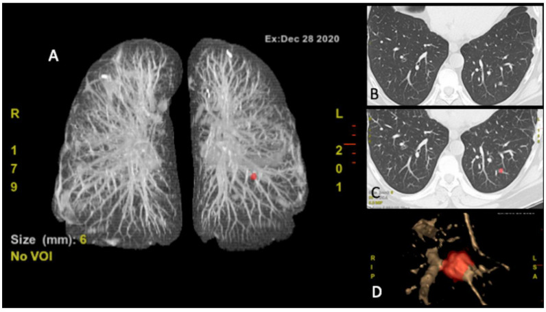
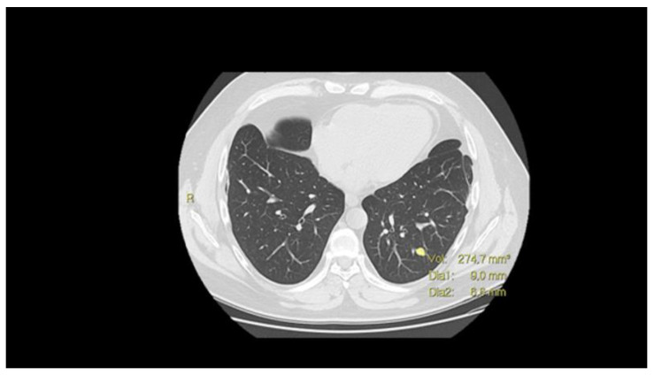
The lung is the most frequent site of osteosarcoma (OS) metastases, which are a critical point in defining a patient's prognosis. Chest computed tomography (CT) represents the gold standard for the detection of lung metastases even if its sensitivity widely ranges in the literature since lung localizations are often atypical. ESMO guidelines represent one of the major references for the follow-up program of OS patients. The development of new reconstruction techniques, such as the iterative method and the deep learning-based image reconstruction (DLIR), has led to a significant reduction of the radiation dose with the low-dose CT. The improvement of these techniques has great importance considering the young-onset of the disease and the strict chest surveillance during follow-up programs. The use of F-fluorodeoxyglucose (FDG) positron emission tomography (PET)/CT is still controversial, while volume doubling time (VDT) and computer-aided diagnosis (CAD) systems are recent diagnostic tools that could support radiologists for lung nodules evaluation. Their use, well-established for other malignancies, needs to be further evaluated, focusing on OS patients.

摘要

肺是骨肉瘤(OS)转移最常见的部位,这是确定患者预后的关键。胸部计算机断层扫描(CT)是检测肺转移的金标准,尽管其敏感性在文献中广泛存在差异,因为肺部定位通常不典型。ESMO 指南是 OS 患者随访计划的主要参考之一。新的重建技术的发展,如迭代方法和基于深度学习的图像重建(DLIR),导致低剂量 CT 的辐射剂量显著降低。考虑到疾病的年轻化和随访计划中严格的胸部监测,这些技术的改进非常重要。氟代脱氧葡萄糖(FDG)正电子发射断层扫描(PET)/CT 的使用仍存在争议,而体积倍增时间(VDT)和计算机辅助诊断(CAD)系统是最近的诊断工具,可以帮助放射科医生评估肺结节。它们在其他恶性肿瘤中的应用已经得到了充分的证实,需要进一步评估,特别是针对 OS 患者。

https://cdn.ncbi.nlm.nih.gov/pmc/blobs/5367/7999261/17b68fce8fde/cells-10-00553-g003.jpg
https://cdn.ncbi.nlm.nih.gov/pmc/blobs/5367/7999261/f9be32cf20b3/cells-10-00553-g001.jpg
https://cdn.ncbi.nlm.nih.gov/pmc/blobs/5367/7999261/979bd2447767/cells-10-00553-g002.jpg
https://cdn.ncbi.nlm.nih.gov/pmc/blobs/5367/7999261/17b68fce8fde/cells-10-00553-g003.jpg
https://cdn.ncbi.nlm.nih.gov/pmc/blobs/5367/7999261/f9be32cf20b3/cells-10-00553-g001.jpg
https://cdn.ncbi.nlm.nih.gov/pmc/blobs/5367/7999261/979bd2447767/cells-10-00553-g002.jpg
https://cdn.ncbi.nlm.nih.gov/pmc/blobs/5367/7999261/17b68fce8fde/cells-10-00553-g003.jpg

相似文献

1
Radiologic Assessment of Osteosarcoma Lung Metastases: State of the Art and Recent Advances.骨 肉 瘤 肺 转 移 的 放 射 学 评 估 : 现 状 和 最 新 进 展 。
Cells. 2021 Mar 4;10(3):553. doi: 10.3390/cells10030553.
2
Computer-aided detection (CAD) of solid pulmonary nodules in chest x-ray equivalent ultralow dose chest CT - first in-vivo results at dose levels of 0.13mSv.胸部X线等效超低剂量胸部CT中实性肺结节的计算机辅助检测——0.13mSv剂量水平下的首次体内研究结果
Eur J Radiol. 2016 Dec;85(12):2217-2224. doi: 10.1016/j.ejrad.2016.10.006. Epub 2016 Oct 11.
3
More advantages in detecting bone and soft tissue metastases from prostate cancer using F-PSMA PET/CT.使用F-PSMA PET/CT检测前列腺癌骨和软组织转移方面有更多优势。
Hell J Nucl Med. 2019 Jan-Apr;22(1):6-9. doi: 10.1967/s002449910952. Epub 2019 Mar 7.
4
Chest radiography or chest CT plus head and neck CT versus F-FDG PET/CT for detection of distant metastasis and synchronous cancer in patients with head and neck cancer.胸部 X 线摄影或胸部 CT 加头颈部 CT 与 F-FDG PET/CT 用于检测头颈部癌症患者的远处转移和同步癌。
Oral Oncol. 2019 Jan;88:109-114. doi: 10.1016/j.oraloncology.2018.11.026. Epub 2018 Nov 23.
5
A cloud-based computer-aided detection system improves identification of lung nodules on computed tomography scans of patients with extra-thoracic malignancies.基于云的计算机辅助检测系统可提高对伴有胸外恶性肿瘤的患者 CT 扫描中肺结节的识别能力。
Eur Radiol. 2019 Jan;29(1):144-152. doi: 10.1007/s00330-018-5528-6. Epub 2018 Jun 15.
6
Pediatric bone sarcoma: diagnostic performance of ¹⁸F-FDG PET/CT versus conventional imaging for initial staging and follow-up.儿童骨肉瘤:¹⁸F-FDG PET/CT 与常规影像学在初始分期和随访中的诊断性能。
AJR Am J Roentgenol. 2015 Jan;204(1):153-60. doi: 10.2214/AJR.14.12932.
7
Prognostic value of metabolic tumor burden on 18F-FDG PET in nonsurgical patients with non-small cell lung cancer.代谢肿瘤负荷 18F-FDG PET 在非手术治疗的非小细胞肺癌患者中的预后价值。
Eur J Nucl Med Mol Imaging. 2012 Jan;39(1):27-38. doi: 10.1007/s00259-011-1934-6. Epub 2011 Sep 23.
8
DaPeCa-7: comparative assessment of fluorodeoxyglucose positron emission tomography/computed tomography (CT) and conventional diagnostic CT in diagnosis of lymph node metastases, distant metastases and incidental findings in patients with invasive penile cancer.DaPeCa-7:氟代脱氧葡萄糖正电子发射断层扫描/计算机断层扫描(CT)与常规诊断 CT 对比评估在浸润性阴茎癌患者淋巴结转移、远处转移和偶然发现中的诊断价值。
BJU Int. 2021 Feb;127(2):254-262. doi: 10.1111/bju.15206. Epub 2020 Sep 7.
9
Assessment of indeterminate pulmonary nodules detected in lung cancer screening: Diagnostic accuracy of FDG PET/CT.肺癌筛查中检测到的肺结节的评估:FDG PET/CT的诊断准确性。
Lung Cancer. 2016 Jul;97:81-6. doi: 10.1016/j.lungcan.2016.04.025. Epub 2016 May 2.
10
Integrating PET and CT information to improve diagnostic accuracy for lung nodules: A semiautomatic computer-aided method.整合正电子发射断层扫描(PET)和计算机断层扫描(CT)信息以提高肺结节诊断准确性:一种半自动计算机辅助方法。
J Nucl Med. 2006 Jul;47(7):1075-80.

引用本文的文献

1
Cluster of differentiation 133 (CD133) and C-X-C chemokine receptor 4 (CXCR4) associated with the incidence of metastasis in osteosarcoma patients.分化簇133(CD133)和C-X-C趋化因子受体4(CXCR4)与骨肉瘤患者的转移发生率相关。
Eur J Orthop Surg Traumatol. 2025 Jul 23;35(1):318. doi: 10.1007/s00590-025-04434-x.
2
Harnessing Artificial Intelligence in Pediatric Oncology Diagnosis and Treatment: A Review.利用人工智能进行儿科肿瘤学诊断与治疗:综述
Cancers (Basel). 2025 May 30;17(11):1828. doi: 10.3390/cancers17111828.
3
New insights into imaging of pulmonary metastases from extra-thoracic neoplasms.

本文引用的文献

1
Incidence Lung Cancer after a Negative CT Screening in the National Lung Screening Trial: Deep Learning-Based Detection of Missed Lung Cancers.国家肺癌筛查试验中CT筛查阴性后的肺癌发病率:基于深度学习的漏诊肺癌检测
J Clin Med. 2020 Dec 2;9(12):3908. doi: 10.3390/jcm9123908.
2
[F]FDG PET/CT for evaluating early response to neoadjuvant chemotherapy in pediatric patients with sarcoma: a prospective single-center trial.[F]氟代脱氧葡萄糖正电子发射断层扫描/计算机断层扫描用于评估小儿肉瘤患者新辅助化疗的早期反应:一项前瞻性单中心试验。
EJNMMI Res. 2020 Oct 15;10(1):122. doi: 10.1186/s13550-020-00715-0.
3
Low-Dose Computed Tomography Screening Proposal for the "Big-3 Diseases": Lung Cancer, Chronic Obstructive Pulmonary Disease, and Cardiovascular Disease.
胸外肿瘤肺转移成像的新见解。
Radiol Med. 2025 Apr 1. doi: 10.1007/s11547-025-02008-9.
4
A Novel Systemic siDR6 Delivery System Based on DP7-C for the Treatment of Metastatic Lung Cancer.一种基于DP7-C的新型系统性siDR6递送系统用于治疗转移性肺癌。
Int J Nanomedicine. 2025 Mar 19;20:3623-3642. doi: 10.2147/IJN.S488213. eCollection 2025.
5
Multimodal Imaging of Osteosarcoma: From First Diagnosis to Radiomics.骨肉瘤的多模态成像:从初次诊断到影像组学
Cancers (Basel). 2025 Feb 10;17(4):599. doi: 10.3390/cancers17040599.
6
Unveiling the Hidden Burden: A Systematic Review on the Prevalence and Clinical Implications of Calcified Brain Metastases.揭示隐藏负担:关于钙化脑转移瘤患病率及临床意义的系统评价
Biomolecules. 2024 Dec 11;14(12):1585. doi: 10.3390/biom14121585.
7
Imaging of Osteosarcoma: Presenting Findings, Metastatic Patterns, and Features Related to Prognosis.骨肉瘤的影像学:呈现的表现、转移模式及与预后相关的特征
J Clin Med. 2024 Sep 25;13(19):5710. doi: 10.3390/jcm13195710.
8
Osteosarcoma Metastasis to the Thorax: A Pictorial Review of Chest Computed Tomography Findings.骨肉瘤转移至胸部:胸部计算机断层扫描结果的影像综述
Diagnostics (Basel). 2024 Sep 20;14(18):2085. doi: 10.3390/diagnostics14182085.
9
In Vivo PET Detection of Lung Micrometastasis in Mice by Targeting Endothelial VCAM-1 Using a Dual-Contrast PET/MRI Probe.利用靶向内皮 VCAM-1 的双对比 PET/MRI 探针在小鼠体内检测肺微转移。
Int J Mol Sci. 2024 Jun 28;25(13):7160. doi: 10.3390/ijms25137160.
10
Predictors of pulmonary metastases on chest computed tomography in children and adolescents with osteosarcoma-tips for qualifying patients for thoracotomy.在儿童和青少年骨肉瘤患者的胸部 CT 上预测肺转移——为开胸手术患者定性的提示。
BMC Pediatr. 2024 Jun 3;24(1):382. doi: 10.1186/s12887-024-04858-0.
针对“三大疾病”(肺癌、慢性阻塞性肺疾病和心血管疾病)的低剂量计算机断层扫描筛查建议
Acad Radiol. 2021 Jan;28(1):46-48. doi: 10.1016/j.acra.2020.07.035. Epub 2020 Aug 15.
4
Validation of Deep-Learning Image Reconstruction for Low-Dose Chest Computed Tomography Scan: Emphasis on Image Quality and Noise.深度学习在低剂量胸部 CT 扫描图像重建中的验证:重点关注图像质量和噪声。
Korean J Radiol. 2021 Jan;22(1):131-138. doi: 10.3348/kjr.2020.0116. Epub 2020 Jul 27.
5
PET with F-Fluorodeoxyglucose/Computed Tomography in the Management of Pediatric Sarcoma.PET 联合 F-Fluorodeoxyglucose/计算机断层扫描在儿科肉瘤治疗中的应用。
PET Clin. 2020 Jul;15(3):333-347. doi: 10.1016/j.cpet.2020.03.008.
6
Radiation Exposure in Pediatric Sarcoma Patients Receiving Initial Curative Chemotherapy.接受初始根治性化疗的小儿肉瘤患者的辐射暴露
J Pediatr Hematol Oncol. 2020 Jan;42(1):e7-e11. doi: 10.1097/MPH.0000000000001677.
7
Deep learning reconstruction improves image quality of abdominal ultra-high-resolution CT.深度学习重建可提高腹部超高分辨率 CT 的图像质量。
Eur Radiol. 2019 Nov;29(11):6163-6171. doi: 10.1007/s00330-019-06170-3. Epub 2019 Apr 11.
8
Risk and clinicopathological features of osteosarcoma metastasis to the lung: A population-based study.骨肉瘤肺转移的风险及临床病理特征:一项基于人群的研究。
J Bone Oncol. 2019 Mar 7;16:100230. doi: 10.1016/j.jbo.2019.100230. eCollection 2019 Jun.
9
Survival and prognosis with osteosarcoma: outcomes in more than 2000 patients in the EURAMOS-1 (European and American Osteosarcoma Study) cohort.骨肉瘤的生存和预后:EURAMOS-1(欧洲和美国骨肉瘤研究)队列中 2000 多例患者的结果。
Eur J Cancer. 2019 Mar;109:36-50. doi: 10.1016/j.ejca.2018.11.027. Epub 2019 Jan 25.
10
Bone sarcomas: ESMO-PaedCan-EURACAN Clinical Practice Guidelines for diagnosis, treatment and follow-up.骨肉瘤:ESMO-儿童癌症-欧洲骨肉瘤协作组诊断、治疗及随访临床实践指南
Ann Oncol. 2018 Oct 1;29(Suppl 4):iv79-iv95. doi: 10.1093/annonc/mdy310.