• 文献检索
  • 文档翻译
  • 深度研究
  • 学术资讯
  • Suppr Zotero 插件Zotero 插件
  • 邀请有礼
  • 套餐&价格
  • 历史记录
应用&插件
Suppr Zotero 插件Zotero 插件浏览器插件Mac 客户端Windows 客户端微信小程序
定价
高级版会员购买积分包购买API积分包
服务
文献检索文档翻译深度研究API 文档MCP 服务
关于我们
关于 Suppr公司介绍联系我们用户协议隐私条款
关注我们

Suppr 超能文献

核心技术专利:CN118964589B侵权必究
粤ICP备2023148730 号-1Suppr @ 2026

文献检索

告别复杂PubMed语法,用中文像聊天一样搜索,搜遍4000万医学文献。AI智能推荐,让科研检索更轻松。

立即免费搜索

文件翻译

保留排版,准确专业,支持PDF/Word/PPT等文件格式,支持 12+语言互译。

免费翻译文档

深度研究

AI帮你快速写综述,25分钟生成高质量综述,智能提取关键信息,辅助科研写作。

立即免费体验

基于石墨烯的光学传感与成像平台中的拉曼和荧光增强方法

Raman and Fluorescence Enhancement Approaches in Graphene-Based Platforms for Optical Sensing and Imaging.

作者信息

Cortijo-Campos Sandra, Ramírez-Jiménez Rafael, de Andrés Alicia

机构信息

Instituto de Ciencia de Materiales de Madrid, Consejo Superior de Investigaciones Científicas, Cantoblanco, 28049 Madrid, Spain.

Departamento de Física, Escuela Politécnica Superior, Universidad Carlos III de Madrid, Avenida Universidad 30, Leganés, 28911 Madrid, Spain.

出版信息

Nanomaterials (Basel). 2021 Mar 5;11(3):644. doi: 10.3390/nano11030644.

DOI:10.3390/nano11030644
PMID:33808013
原文链接:https://pmc.ncbi.nlm.nih.gov/articles/PMC7999291/
Abstract

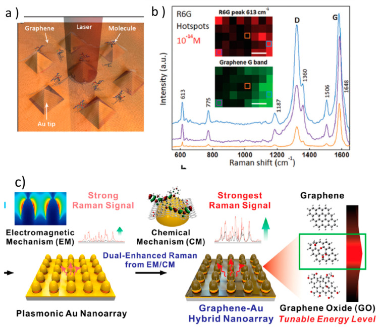
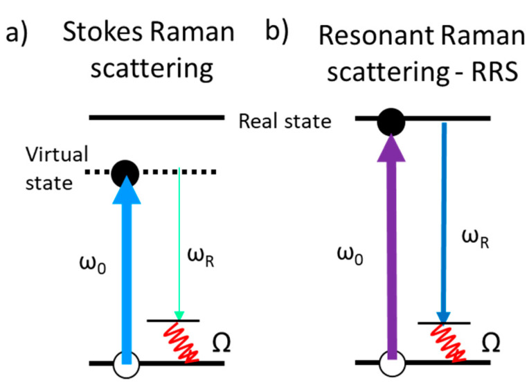
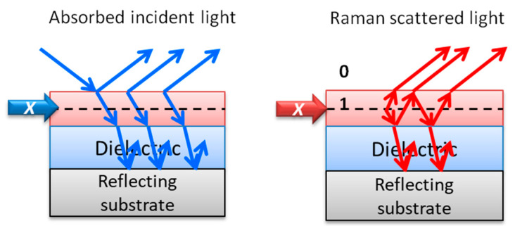
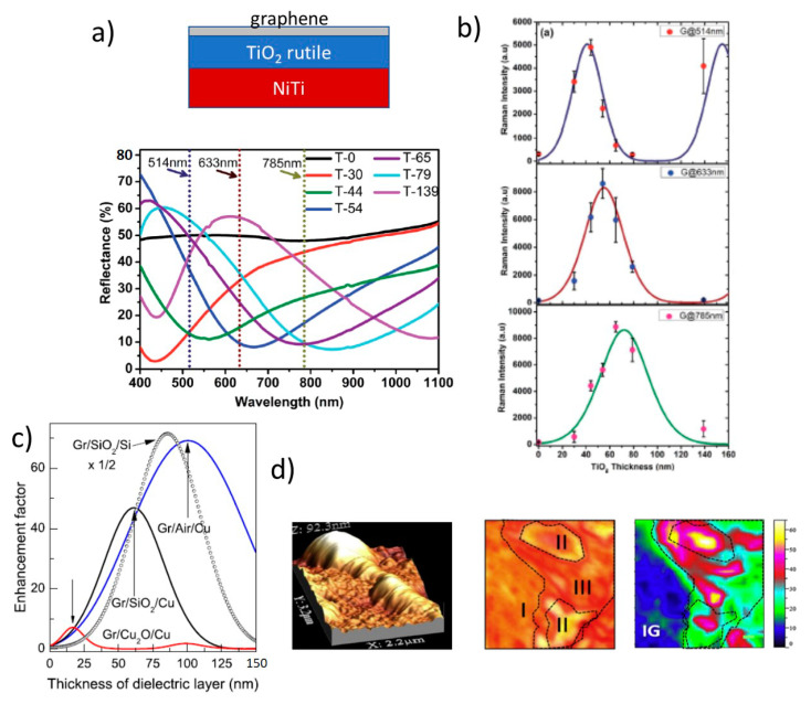
The search for novel platforms and metamaterials for the enhancement of optical and particularly Raman signals is still an objective since optical techniques offer affordable, noninvasive methods with high spatial resolution and penetration depth adequate to detect and image a large variety of systems, from 2D materials to molecules in complex media and tissues. Definitely, plasmonic materials produce the most efficient enhancement through the surface-enhanced Raman scattering (SERS) process, allowing single-molecule detection, and are the most studied ones. Here we focus on less explored aspects of SERS such as the role of the inter-nanoparticle (NP) distance and the ultra-small NP size limit (down to a few nm) and on novel approaches involving graphene and graphene-related materials. The issues on reproducibility and homogeneity for the quantification of the probe molecules will also be discussed. Other light enhancement mechanisms, in particular resonant and interference Raman scatterings, as well as the platforms that allow combining several of them, are presented in this review with a special focus on the possibilities that graphene offers for the design and fabrication of novel architectures. Recent fluorescence enhancement platforms and strategies, so important for bio-detection and imaging, are reviewed as well as the relevance of graphene oxide and graphene/carbon nanodots in the field.

摘要

寻找用于增强光学信号尤其是拉曼信号的新型平台和超材料仍然是一个目标,因为光学技术提供了经济实惠、非侵入性的方法,具有高空间分辨率和足够的穿透深度,足以检测和成像从二维材料到复杂介质和组织中的分子等各种各样的系统。毫无疑问,等离子体材料通过表面增强拉曼散射(SERS)过程产生最有效的增强效果,可实现单分子检测,并且是研究最多的材料。在此,我们关注SERS较少被探索的方面,例如纳米颗粒间距离的作用和超小纳米颗粒尺寸极限(低至几纳米),以及涉及石墨烯和石墨烯相关材料的新方法。还将讨论探针分子定量中的重现性和均匀性问题。本综述介绍了其他光增强机制,特别是共振拉曼散射和干涉拉曼散射,以及能够将其中几种机制结合起来的平台,并特别关注石墨烯为新型结构的设计和制造所提供的可能性。还综述了近期对生物检测和成像非常重要的荧光增强平台和策略,以及氧化石墨烯和石墨烯/碳纳米点在该领域的相关性。

https://cdn.ncbi.nlm.nih.gov/pmc/blobs/481c/7999291/6f6b3bd383b9/nanomaterials-11-00644-g018.jpg
https://cdn.ncbi.nlm.nih.gov/pmc/blobs/481c/7999291/2c5983f12cdc/nanomaterials-11-00644-g001.jpg
https://cdn.ncbi.nlm.nih.gov/pmc/blobs/481c/7999291/800de0866222/nanomaterials-11-00644-g002.jpg
https://cdn.ncbi.nlm.nih.gov/pmc/blobs/481c/7999291/ff1b0e364fb2/nanomaterials-11-00644-g003.jpg
https://cdn.ncbi.nlm.nih.gov/pmc/blobs/481c/7999291/07484cf561ce/nanomaterials-11-00644-g004.jpg
https://cdn.ncbi.nlm.nih.gov/pmc/blobs/481c/7999291/e6e8ce963056/nanomaterials-11-00644-g005.jpg
https://cdn.ncbi.nlm.nih.gov/pmc/blobs/481c/7999291/ee331f6de8eb/nanomaterials-11-00644-g006.jpg
https://cdn.ncbi.nlm.nih.gov/pmc/blobs/481c/7999291/0f95719d2db2/nanomaterials-11-00644-g007.jpg
https://cdn.ncbi.nlm.nih.gov/pmc/blobs/481c/7999291/d7b3591c224e/nanomaterials-11-00644-g008.jpg
https://cdn.ncbi.nlm.nih.gov/pmc/blobs/481c/7999291/5a4e520fdda7/nanomaterials-11-00644-g009.jpg
https://cdn.ncbi.nlm.nih.gov/pmc/blobs/481c/7999291/63414ab6deed/nanomaterials-11-00644-g010.jpg
https://cdn.ncbi.nlm.nih.gov/pmc/blobs/481c/7999291/f4996a22f9df/nanomaterials-11-00644-g011.jpg
https://cdn.ncbi.nlm.nih.gov/pmc/blobs/481c/7999291/5dde4aa335bb/nanomaterials-11-00644-g012.jpg
https://cdn.ncbi.nlm.nih.gov/pmc/blobs/481c/7999291/a4c4dcf3fbbe/nanomaterials-11-00644-g013.jpg
https://cdn.ncbi.nlm.nih.gov/pmc/blobs/481c/7999291/716a2c604869/nanomaterials-11-00644-g014.jpg
https://cdn.ncbi.nlm.nih.gov/pmc/blobs/481c/7999291/5691e8f57ca3/nanomaterials-11-00644-g015.jpg
https://cdn.ncbi.nlm.nih.gov/pmc/blobs/481c/7999291/b1280883d333/nanomaterials-11-00644-g016.jpg
https://cdn.ncbi.nlm.nih.gov/pmc/blobs/481c/7999291/8555527485aa/nanomaterials-11-00644-g017.jpg
https://cdn.ncbi.nlm.nih.gov/pmc/blobs/481c/7999291/6f6b3bd383b9/nanomaterials-11-00644-g018.jpg

相似文献

1
Raman and Fluorescence Enhancement Approaches in Graphene-Based Platforms for Optical Sensing and Imaging.基于石墨烯的光学传感与成像平台中的拉曼和荧光增强方法
Nanomaterials (Basel). 2021 Mar 5;11(3):644. doi: 10.3390/nano11030644.
2
Supported Ultra-Thin Alumina Membranes with Graphene as Efficient Interference Enhanced Raman Scattering Platforms for Sensing.以石墨烯为高效干涉增强拉曼散射传感平台的负载型超薄氧化铝膜
Nanomaterials (Basel). 2020 Apr 27;10(5):830. doi: 10.3390/nano10050830.
3
Efficient Heterostructures for Combined Interference and Plasmon Resonance Raman Amplification.用于干涉和等离子体共振拉曼放大组合的高效异质结构。
ACS Appl Mater Interfaces. 2017 Feb 1;9(4):4119-4125. doi: 10.1021/acsami.6b12490. Epub 2017 Jan 18.
4
Dual-Enhanced Raman Scattering-Based Characterization of Stem Cell Differentiation Using Graphene-Plasmonic Hybrid Nanoarray.基于石墨烯等离子体混合纳阵列的双重增强拉曼散射对干细胞分化的特性分析。
Nano Lett. 2019 Nov 13;19(11):8138-8148. doi: 10.1021/acs.nanolett.9b03402. Epub 2019 Nov 1.
5
Controlled Electrodeposition of Gold on Graphene: Maximization of the Defect-Enhanced Raman Scattering Response.金在石墨烯上的可控电沉积:最大化缺陷增强拉曼散射响应。
Small. 2019 Nov;15(48):e1901555. doi: 10.1002/smll.201901555. Epub 2019 May 21.
6
Strong Dependence of Surface Enhanced Raman Scattering on Structure of Graphene Oxide Film.表面增强拉曼散射对氧化石墨烯薄膜结构的强烈依赖性。
Materials (Basel). 2018 Jul 12;11(7):1199. doi: 10.3390/ma11071199.
7
Graphene oxide and gold nanoparticle based dual platform with short DNA probe for the PCR free DNA biosensing using surface-enhanced Raman scattering.基于氧化石墨烯和金纳米粒子的双平台,结合短 DNA 探针,实现了无需聚合酶链反应的 DNA 生物传感,利用表面增强拉曼散射。
Biosens Bioelectron. 2019 Apr 15;131:214-223. doi: 10.1016/j.bios.2019.02.028. Epub 2019 Feb 19.
8
Plasmonic Nanogap-Enhanced Raman Scattering with Nanoparticles.等离子体纳米间隙增强拉曼散射与纳米粒子。
Acc Chem Res. 2016 Dec 20;49(12):2746-2755. doi: 10.1021/acs.accounts.6b00409. Epub 2016 Nov 8.
9
Designing surface-enhanced Raman scattering (SERS) platforms beyond hotspot engineering: emerging opportunities in analyte manipulations and hybrid materials.超越热点工程的设计:表面增强拉曼散射 (SERS) 平台中的新兴分析物操控和混合材料机遇。
Chem Soc Rev. 2019 Feb 4;48(3):731-756. doi: 10.1039/c7cs00786h.
10
Strong light-matter interactions in sub-nanometer gaps defined by monolayer graphene: toward highly sensitive SERS substrates.由单层石墨烯定义的亚纳米间隙中的强光物质相互作用:迈向高灵敏度 SERS 衬底。
Nanoscale. 2014 Oct 7;6(19):11112-20. doi: 10.1039/c4nr03152k.

引用本文的文献

1
UV-promoted surface-enhanced Raman spectroscopy via heterojunction of few-layer MoS flakes on acceptor-rich β-GaO microstrips.通过在富含受体的β-Ga₂O₃微带上的少层MoS₂薄片异质结实现紫外线促进的表面增强拉曼光谱
Discov Nano. 2025 Sep 5;20(1):152. doi: 10.1186/s11671-025-04339-y.
2
Fluorescence Based Trace Level Detection of Cobalt Using 2,6-bis((E)-4-chlorobenzylidene)-4-methylcyclohexan-1-one as Sensor.以2,6-双((E)-4-氯亚苄基)-4-甲基环己酮为传感器基于荧光的痕量钴检测
J Fluoresc. 2025 Apr 23. doi: 10.1007/s10895-025-04306-x.
3
Advances in the application of Raman spectroscopy in haematological tumours.

本文引用的文献

1
Raman amplification in the ultra-small limit of Ag nanoparticles on SiO and graphene: Size and inter-particle distance effects.SiO和石墨烯上银纳米颗粒超小尺寸极限下的拉曼放大:尺寸和颗粒间距离效应。
Mater Des. 2020 Apr 2;192:108702. doi: 10.1016/j.matdes.2020.108702. eCollection 2020 Jul.
2
Real-time detection of single-molecule reaction by plasmon-enhanced spectroscopy.通过等离子体增强光谱法实时检测单分子反应。
Sci Adv. 2020 Jun 10;6(24):eaba6012. doi: 10.1126/sciadv.aba6012. eCollection 2020 Jun.
3
Mapping Fluorescence Enhancement of Plasmonic Nanorod Coupled Dye Molecules.
拉曼光谱在血液系统肿瘤中的应用进展
Front Bioeng Biotechnol. 2023 Jan 10;10:1103785. doi: 10.3389/fbioe.2022.1103785. eCollection 2022.
4
One-Step Homogeneous Immunoassay for the Detection of Influenza Virus Using Switching Peptide and Graphene Quencher.基于开关肽和石墨烯猝灭剂的一步均相免疫分析法检测流感病毒
Biochip J. 2022;16(3):334-341. doi: 10.1007/s13206-022-00076-x. Epub 2022 Jul 27.
绘制等离子体纳米棒耦合染料分子的荧光增强图谱。
Nanomaterials (Basel). 2020 May 29;10(6):1048. doi: 10.3390/nano10061048.
4
Supported Ultra-Thin Alumina Membranes with Graphene as Efficient Interference Enhanced Raman Scattering Platforms for Sensing.以石墨烯为高效干涉增强拉曼散射传感平台的负载型超薄氧化铝膜
Nanomaterials (Basel). 2020 Apr 27;10(5):830. doi: 10.3390/nano10050830.
5
Surface-Enhanced Raman Spectroscopy on Hybrid Graphene/Gold Substrates near the Percolation Threshold.渗流阈值附近混合石墨烯/金基底上的表面增强拉曼光谱
Nanomaterials (Basel). 2020 Jan 17;10(1):164. doi: 10.3390/nano10010164.
6
Galvanic Replacement Reaction as a Route to Prepare Nanoporous Aluminum for UV Plasmonics.通过电偶置换反应制备用于紫外等离子体的纳米多孔铝
Nanomaterials (Basel). 2020 Jan 4;10(1):102. doi: 10.3390/nano10010102.
7
Dual-Enhanced Raman Scattering-Based Characterization of Stem Cell Differentiation Using Graphene-Plasmonic Hybrid Nanoarray.基于石墨烯等离子体混合纳阵列的双重增强拉曼散射对干细胞分化的特性分析。
Nano Lett. 2019 Nov 13;19(11):8138-8148. doi: 10.1021/acs.nanolett.9b03402. Epub 2019 Nov 1.
8
Ultrabright gap-enhanced Raman tags for high-speed bioimaging.超亮间隙增强拉曼标签用于高速生物成像。
Nat Commun. 2019 Aug 29;10(1):3905. doi: 10.1038/s41467-019-11829-y.
9
Plasmon-Enhanced Fluorescence of Carbon Nanodots in Gold Nanoslit Cavities.金纳米狭缝腔中碳纳米点的表面等离子体增强荧光
Langmuir. 2019 Jul 9;35(27):8903-8909. doi: 10.1021/acs.langmuir.9b00448. Epub 2019 Jun 27.
10
Chemical and Bio Sensing Using Graphene-Enhanced Raman Spectroscopy.利用石墨烯增强拉曼光谱的化学与生物传感
Nanomaterials (Basel). 2019 Apr 2;9(4):516. doi: 10.3390/nano9040516.