Takao S, Miyatake K, Izumi S, Okamoto M, Kinoshita N, Nakagawa H, Yamamoto K, Sakakibara H, Nimura Y
Cardiology Division, National Cardiovascular Centre, Osaka, Japan.
Br Heart J. 1988 May;59(5):542-50. doi: 10.1136/hrt.59.5.542.
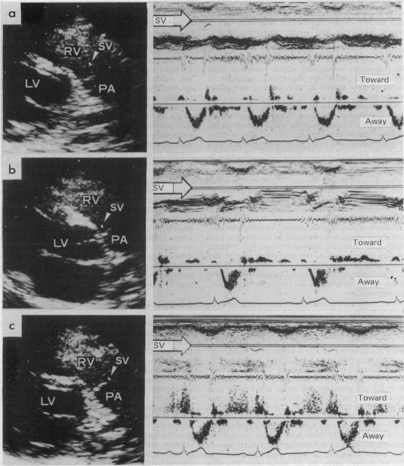
Pulsed Doppler echocardiography in healthy individuals often shows a disturbance of diastolic flow in the right ventricular outflow tract just below the pulmonary valve that suggests regurgitation. This disturbance of diastolic flow was studied in 50 healthy individuals and 40 patients with cardiopulmonary disease, some of whom had a pulmonary regurgitant murmur. Diastolic flow was disturbed in 39 of the 50 healthy individuals. In 32, cross sectional echocardiography gave a satisfactory image of the pulmonary valve. The characteristic Doppler signals usually lasted throughout diastole, were directed toward the right ventricular cavity, and gradually waned towards end diastole; they formed a spindle shaped area of abnormal signals that extended to within 10 mm of the coaptation of the pulmonary valve towards the right ventricular cavity and the pressure difference estimated from the signals by the modified Bernoulli equation seemed to be proportional to the normal retrograde transpulmonary pressure difference. In all 40 patients with cardiopulmonary disease, signals indicating pulmonary regurgitation were found whether or not a regurgitant murmur was present. When it was present, however, the spindle was longer than 20 mm and in patients with pulmonary hypertension the velocity of abnormal diastolic flow was higher than in healthy individuals. The Doppler signals registering disturbed flow in the healthy individuals resembled the signals caused by pulmonary regurgitation in the patients in terms of location, orientation, and configuration. These results show that healthy individuals usually have trivial pulmonary regurgitation. In practice the distance that the flow disturbance extends from the valve and estimated pressure difference across the valve are probably the most important variables for assessing the clinical significance of pulmonary valve regurgitation.
健康个体的脉冲多普勒超声心动图常显示肺动脉瓣下方右心室流出道舒张期血流紊乱,提示存在反流。对50名健康个体和40名心肺疾病患者的这种舒张期血流紊乱情况进行了研究,其中一些患者有肺动脉反流杂音。50名健康个体中有39名存在舒张期血流紊乱。32名个体的横截面超声心动图能清晰显示肺动脉瓣。特征性多普勒信号通常在整个舒张期持续存在,信号指向右心室腔,在舒张期末期逐渐减弱;它们形成一个纺锤形异常信号区,延伸至肺动脉瓣与右心室腔贴合处10毫米范围内,通过改良伯努利方程根据信号估算的压力差似乎与正常的逆行跨肺压力差成正比。在所有40名心肺疾病患者中,无论是否存在反流杂音,均发现了提示肺动脉反流的信号。然而,当存在反流杂音时,纺锤形信号区长度超过20毫米,在肺动脉高压患者中,舒张期异常血流速度高于健康个体。健康个体中记录到的血流紊乱的多普勒信号在位置、方向和形态上与患者肺动脉反流产生的信号相似。这些结果表明,健康个体通常存在轻微的肺动脉反流。在实际应用中,血流紊乱从瓣膜延伸的距离以及估算的跨瓣膜压力差可能是评估肺动脉瓣反流临床意义的最重要变量。