Department of Molecular Biosciences, Rice Institute for Biomedical Research, Northwestern University, Evanston, IL 60208-3500;
Centre for Misfolding Diseases, Yusuf Hamied Department of Chemistry, University of Cambridge, CB2 1EW Cambridge, United Kingdom.
Proc Natl Acad Sci U S A. 2021 Mar 16;118(11). doi: 10.1073/pnas.2021888118.
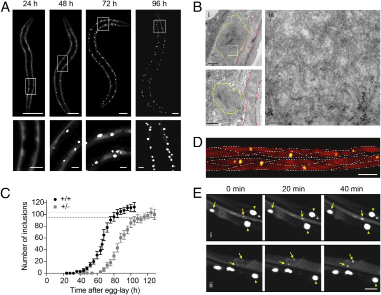
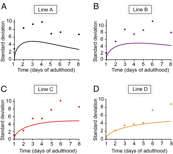
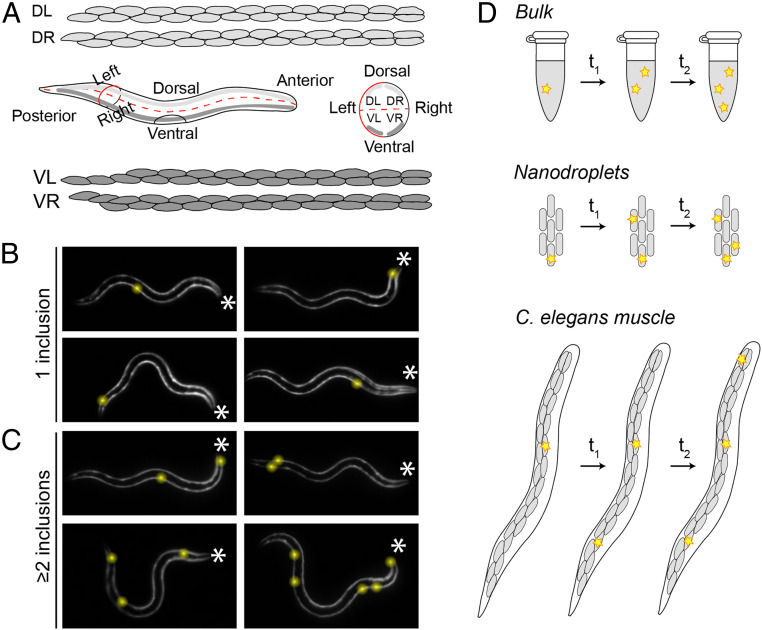
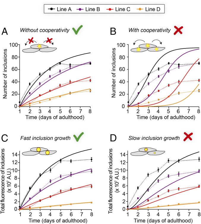
Protein aggregation is associated with a wide range of degenerative human diseases with devastating consequences, as exemplified by Alzheimer's, Parkinson's, and Huntington's diseases. In vitro kinetic studies have provided a mechanistic understanding of the aggregation process at the molecular level. However, it has so far remained largely unclear to what extent the biophysical principles of amyloid formation learned in vitro translate to the complex environment of living organisms. Here, we take advantage of the unique properties of a model expressing a fluorescently tagged polyglutamine (polyQ) protein, which aggregates into discrete micrometer-sized inclusions that can be directly visualized in real time. We provide a quantitative analysis of protein aggregation in this system and show that the data are described by a molecular model where stochastic nucleation occurs independently in each cell, followed by rapid aggregate growth. Global fitting of the image-based aggregation kinetics reveals a nucleation rate corresponding to 0.01 h per cell at 1 mM intracellular protein concentration, and shows that the intrinsic molecular stochasticity of nucleation accounts for a significant fraction of the observed animal-to-animal variation. Our results highlight how independent, stochastic nucleation events in individual cells control the overall progression of polyQ aggregation in a living animal. The key finding that the biophysical principles associated with protein aggregation in small volumes remain the governing factors, even in the complex environment of a living organism, will be critical for the interpretation of in vivo data from a wide range of protein aggregation diseases.
蛋白质聚集与广泛的退行性人类疾病有关,这些疾病具有破坏性的后果,如阿尔茨海默病、帕金森病和亨廷顿病。体外动力学研究提供了在分子水平上对聚集过程的机制理解。然而,到目前为止,在多大程度上,体外获得的淀粉样蛋白形成的生物物理原理转化为生物体的复杂环境,仍然很大程度上不清楚。在这里,我们利用表达荧光标记的多聚谷氨酰胺(polyQ)蛋白的模型的独特性质,该蛋白聚集成离散的微米大小的包含物,可以实时直接可视化。我们对该系统中的蛋白质聚集进行了定量分析,并表明数据可以用一个分子模型来描述,其中随机成核在每个细胞中独立发生,然后快速进行聚集体生长。基于图像的聚集动力学的全局拟合揭示了在 1 mM 细胞内蛋白质浓度下,每个细胞的成核速率为 0.01 h,并且表明成核的固有分子随机性解释了观察到的动物间变异性的很大一部分。我们的结果强调了单个细胞中独立的、随机的成核事件如何控制聚 Q 聚集在活体动物中的整体进展。关键的发现是,与小体积中的蛋白质聚集相关的生物物理原理仍然是控制因素,即使在生物体的复杂环境中也是如此,这对于解释广泛的蛋白质聚集疾病的体内数据将是至关重要的。