• 文献检索
  • 文档翻译
  • 深度研究
  • 学术资讯
  • Suppr Zotero 插件Zotero 插件
  • 邀请有礼
  • 套餐&价格
  • 历史记录
应用&插件
Suppr Zotero 插件Zotero 插件浏览器插件Mac 客户端Windows 客户端微信小程序
定价
高级版会员购买积分包购买API积分包
服务
文献检索文档翻译深度研究API 文档MCP 服务
关于我们
关于 Suppr公司介绍联系我们用户协议隐私条款
关注我们

Suppr 超能文献

核心技术专利:CN118964589B侵权必究
粤ICP备2023148730 号-1Suppr @ 2026

文献检索

告别复杂PubMed语法,用中文像聊天一样搜索,搜遍4000万医学文献。AI智能推荐,让科研检索更轻松。

立即免费搜索

文件翻译

保留排版,准确专业,支持PDF/Word/PPT等文件格式,支持 12+语言互译。

免费翻译文档

深度研究

AI帮你快速写综述,25分钟生成高质量综述,智能提取关键信息,辅助科研写作。

立即免费体验

胎儿极早早产儿的脑容量测量:一项 MRI 初步研究。

Brain volumetry in fetuses that deliver very preterm: An MRI pilot study.

机构信息

Department of Women and Children's Health, King's College London, UK.

Centre for the Developing Brain, King's College London, London, UK.

出版信息

Neuroimage Clin. 2021;30:102650. doi: 10.1016/j.nicl.2021.102650. Epub 2021 Mar 29.

DOI:10.1016/j.nicl.2021.102650
PMID:33838546
原文链接:https://pmc.ncbi.nlm.nih.gov/articles/PMC8045030/
Abstract

BACKGROUND

Infants born preterm are at increased risk of neurological complications resulting in significant morbidity and mortality. The exact mechanism and the impact of antenatal factors has not been fully elucidated, although antenatal infection/inflammation has been implicated in both the aetiology of preterm birth and subsequent neurological sequelae. It is therefore hypothesized that processes driving preterm birth are affecting brain development in utero. This study aims to compare MRI derived regional brain volumes in fetuses that deliver < 32 weeks with fetuses that subsequently deliver at term.

METHODS

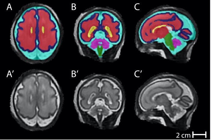
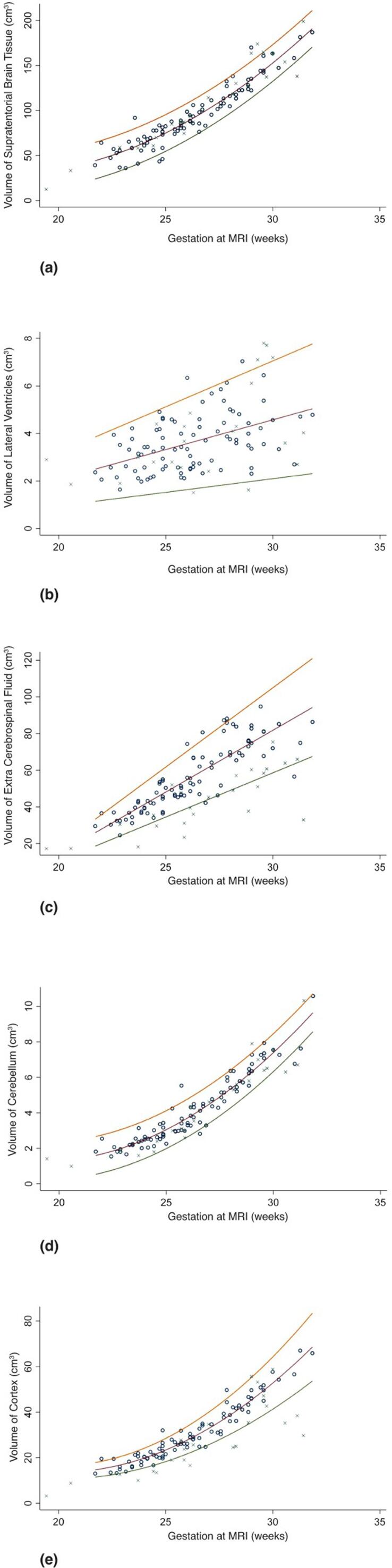
Women at high risk of preterm birth, with gestation 19.4-32 weeks were recruited prospectively. A control group was obtained from existing study datasets. Fetal MRI was performed on a 1.5 T or 3 T MRI scanner: T2-weighted images were obtained of the fetal brain. 3D brain volumetric datsets were produced using slice to volume reconstruction and regional segmentations were produced using multi-atlas approaches for supratentorial brain tissue, lateral ventricles, cerebellum cerebral cortex and extra-cerebrospinal fluid (eCSF). Statistical comparison of control and high-risk for preterm delivery fetuses was performed by creating normal ranges for each parameter from the control datasets and then calculating gestation adjusted z scores. Groups were compared using t-tests.

RESULTS

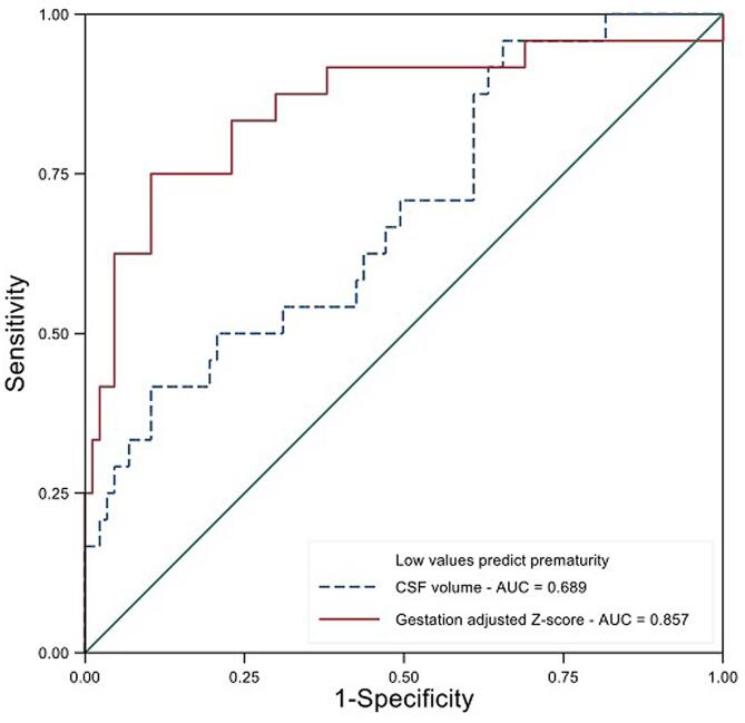
Fetal image datasets from 24 pregnancies with delivery < 32 weeks and 87 control pregnancies that delivered > 37 weeks were included. Median gestation at MRI of the preterm group was 26.8 weeks (range 19.4-31.4) and control group 26.2 weeks (range 21.7-31.9). No difference was found in supra-tentorial brain volume, ventricular volume or cerebellar volume but the eCSF and cerebral cortex volumes were smaller in fetuses that delivered preterm (p < 0.001 in both cases).

CONCLUSION

Fetuses that deliver preterm have a reduction in cortical and eCSF volumes. This is a novel finding and needs further investigation. If alterations in brain development are commencing antenatally in fetuses that subsequently deliver preterm, this may present a window for in utero therapy in the future.

摘要

背景

早产儿存在神经并发症风险增加的情况,这导致了较高的发病率和死亡率。尽管产前感染/炎症与早产的病因以及随后的神经后遗症都有关,但确切的机制和产前因素的影响尚未完全阐明。因此,有人假设导致早产的过程正在子宫内影响大脑发育。本研究旨在比较<32 周分娩的胎儿与随后足月分娩的胎儿的 MRI 得出的区域性脑容量。

方法

高风险早产的孕妇,妊娠 19.4-32 周,前瞻性招募。对照组从现有研究数据集获得。对胎儿进行 1.5T 或 3T MRI 扫描:获得胎儿大脑的 T2 加权图像。使用切片到体积重建获得 3D 脑容积数据集,并使用多图谱方法对幕上脑组织、侧脑室、小脑、大脑皮层和额外脑脊髓液(eCSF)进行区域分割。通过为对照组数据集创建每个参数的正常范围,然后计算妊娠调整 z 分数,对控制组和早产高风险胎儿进行统计比较。使用 t 检验比较组间差异。

结果

共纳入 24 例<32 周分娩和 87 例>37 周分娩的胎儿的胎儿图像数据集。早产组 MRI 的中位妊娠周数为 26.8 周(范围 19.4-31.4),对照组为 26.2 周(范围 21.7-31.9)。幕上脑容量、脑室容量或小脑容量无差异,但早产儿脑室 eCSF 和大脑皮层体积较小(均 p<0.001)。

结论

早产儿的皮质和 eCSF 体积减少。这是一个新的发现,需要进一步研究。如果在随后早产的胎儿中,脑发育的改变在产前就已经开始,那么这可能为未来的宫内治疗提供了一个窗口。

https://cdn.ncbi.nlm.nih.gov/pmc/blobs/db7d/8045030/e2908a5318da/gr3.jpg
https://cdn.ncbi.nlm.nih.gov/pmc/blobs/db7d/8045030/c467260280ad/gr1.jpg
https://cdn.ncbi.nlm.nih.gov/pmc/blobs/db7d/8045030/92b0f126497e/gr2.jpg
https://cdn.ncbi.nlm.nih.gov/pmc/blobs/db7d/8045030/e2908a5318da/gr3.jpg
https://cdn.ncbi.nlm.nih.gov/pmc/blobs/db7d/8045030/c467260280ad/gr1.jpg
https://cdn.ncbi.nlm.nih.gov/pmc/blobs/db7d/8045030/92b0f126497e/gr2.jpg
https://cdn.ncbi.nlm.nih.gov/pmc/blobs/db7d/8045030/e2908a5318da/gr3.jpg

相似文献

1
Brain volumetry in fetuses that deliver very preterm: An MRI pilot study.胎儿极早早产儿的脑容量测量:一项 MRI 初步研究。
Neuroimage Clin. 2021;30:102650. doi: 10.1016/j.nicl.2021.102650. Epub 2021 Mar 29.
2
Antenatal thymus volumes in fetuses that delivered <32 weeks' gestation: An MRI pilot study.胎儿在 32 周前分娩的产前胸腺体积:一项 MRI 初步研究。
Acta Obstet Gynecol Scand. 2021 Jun;100(6):1040-1050. doi: 10.1111/aogs.13983. Epub 2020 Sep 24.
3
Functional MRI assessment of the lungs in fetuses that deliver very Preterm: An MRI pilot study.功能磁共振成像评估极早产儿肺功能:一项 MRI 初步研究。
Eur J Obstet Gynecol Reprod Biol. 2024 Feb;293:106-114. doi: 10.1016/j.ejogrb.2023.12.015. Epub 2023 Dec 20.
4
Adrenal volumes in fetuses delivering prior to 32 weeks' gestation: An MRI pilot study.胎儿在 32 周前分娩时的肾上腺体积:一项 MRI 初步研究。
Acta Obstet Gynecol Scand. 2024 Mar;103(3):512-521. doi: 10.1111/aogs.14733. Epub 2023 Nov 27.
5
Functional assessment of brain development in fetuses that subsequently deliver very preterm: An MRI pilot study.胎儿期大脑发育的功能评估:随后早产的 MRI 初步研究。
Prenat Diagn. 2024 Jan;44(1):49-56. doi: 10.1002/pd.6498. Epub 2023 Dec 21.
6
Third Trimester Brain Growth in Preterm Infants Compared With In Utero Healthy Fetuses.与宫内健康胎儿相比,早产儿孕晚期的脑发育情况
Pediatrics. 2016 Nov;138(5). doi: 10.1542/peds.2016-1640.
7
Successful use of an artificial placenta-based life support system to treat extremely preterm ovine fetuses compromised by intrauterine inflammation.成功使用基于人工胎盘的生命支持系统治疗因宫内炎症而受损的极早产儿羊胎。
Am J Obstet Gynecol. 2020 Nov;223(5):755.e1-755.e20. doi: 10.1016/j.ajog.2020.04.036. Epub 2020 May 4.
8
Foetal lung volumes in pregnant women who deliver very preterm: a pilot study.早产儿孕妇胎儿肺容积:一项初步研究。
Pediatr Res. 2020 May;87(6):1066-1071. doi: 10.1038/s41390-019-0717-9. Epub 2019 Dec 7.
9
T2*-weighted placental magnetic resonance imaging: a biomarker of placental dysfunction in small-for-gestational-age pregnancies.T2*-加权胎盘磁共振成像:胎儿生长受限妊娠中胎盘功能障碍的生物标志物。
Am J Obstet Gynecol MFM. 2022 May;4(3):100578. doi: 10.1016/j.ajogmf.2022.100578. Epub 2022 Jan 31.
10
Brainstem and cerebellar volumes at magnetic resonance imaging are smaller in fetuses with congenital heart disease.磁共振成像显示,先天性心脏病胎儿的脑干和小脑体积较小。
Am J Obstet Gynecol. 2022 Aug;227(2):282.e1-282.e15. doi: 10.1016/j.ajog.2022.03.030. Epub 2022 Mar 16.

引用本文的文献

1
Burst of gyrification in the human brain after birth.出生后人类大脑脑回形成的爆发期。
Commun Biol. 2025 May 26;8(1):805. doi: 10.1038/s42003-025-08155-z.
2
Scanner-based real-time three-dimensional brain + body slice-to-volume reconstruction for T2-weighted 0.55-T low-field fetal magnetic resonance imaging.基于扫描仪的实时三维脑+体层切片到体积重建,用于0.55-T低场T2加权胎儿磁共振成像。
Pediatr Radiol. 2025 Mar;55(3):556-569. doi: 10.1007/s00247-025-06165-x. Epub 2025 Jan 24.
3
T* relaxometry of fetal brain structures using low-field (0.55T) MRI.
使用低场(0.55T)MRI对胎儿脑结构进行T* 弛豫测量法。
Magn Reson Med. 2025 May;93(5):1942-1953. doi: 10.1002/mrm.30409. Epub 2024 Dec 31.
4
Craniofacial phenotyping with fetal MRI: a feasibility study of 3D visualisation, segmentation, surface-rendered and physical models.胎儿 MRI 的颅面表型分析:3D 可视化、分割、表面渲染和物理模型的可行性研究。
BMC Med Imaging. 2024 Mar 1;24(1):52. doi: 10.1186/s12880-024-01230-7.
5
Functional assessment of brain development in fetuses that subsequently deliver very preterm: An MRI pilot study.胎儿期大脑发育的功能评估:随后早产的 MRI 初步研究。
Prenat Diagn. 2024 Jan;44(1):49-56. doi: 10.1002/pd.6498. Epub 2023 Dec 21.
6
Adrenal volumes in fetuses delivering prior to 32 weeks' gestation: An MRI pilot study.胎儿在 32 周前分娩时的肾上腺体积:一项 MRI 初步研究。
Acta Obstet Gynecol Scand. 2024 Mar;103(3):512-521. doi: 10.1111/aogs.14733. Epub 2023 Nov 27.
7
Mechanical ventilation induces brainstem inflammation in preterm fetal sheep.机械通气可诱发早产胎羊脑干炎症。
Front Pediatr. 2023 Oct 23;11:1225294. doi: 10.3389/fped.2023.1225294. eCollection 2023.
8
Key roles of glial cells in the encephalopathy of prematurity.星形胶质细胞在早产儿脑病中的关键作用。
Glia. 2024 Mar;72(3):475-503. doi: 10.1002/glia.24474. Epub 2023 Nov 1.
9
Reliability and Feasibility of Low-Field-Strength Fetal MRI at 0.55 T during Pregnancy.0.55T 低磁场强度胎儿 MRI 在妊娠期的可靠性和可行性。
Radiology. 2023 Oct;309(1):e223050. doi: 10.1148/radiol.223050.
10
BOUNTI: Brain vOlumetry and aUtomated parcellatioN for 3D feTal MRI.BOUNTI:用于三维胎儿磁共振成像的脑容量测定与自动脑区划分
bioRxiv. 2023 Apr 27:2023.04.18.537347. doi: 10.1101/2023.04.18.537347.