Payette Kelly, Uus Alena U, Kollstad Ella, Aviles Verdera Jordina, Gallo Dario, Hall Megan, Hajnal Joseph V, Rutherford Mary A, Story Lisa, Hutter Jana
Research Department of Early Life Imaging, School of Biomedical Engineering and Imaging Sciences, King's College London, London, UK.
Biomedical Engineering Department, School of Biomedical Engineering and Imaging Sciences, King's College London, London, UK.
Magn Reson Med. 2025 May;93(5):1942-1953. doi: 10.1002/mrm.30409. Epub 2024 Dec 31.
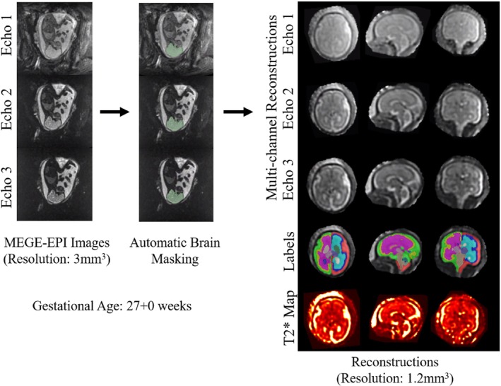
Human brain development during gestation is complex, as both structure and function are rapidly forming. Structural imaging methods using MRI are well developed to explore these changes, but functional imaging tools are lacking. Low-field MRI is a promising modality to bridge this gap. The longer intrinsic T* values at low field strengths increase the dynamic range and enable the quantification of individual brain regions with low T* values, such as deep gray matter. This study investigates regional brain T* quantification throughout the second half of gestation on low-field 0.55T MRI.
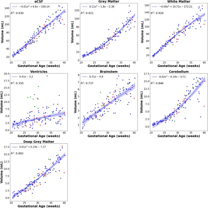
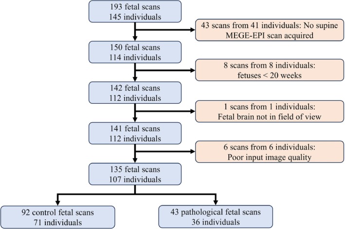
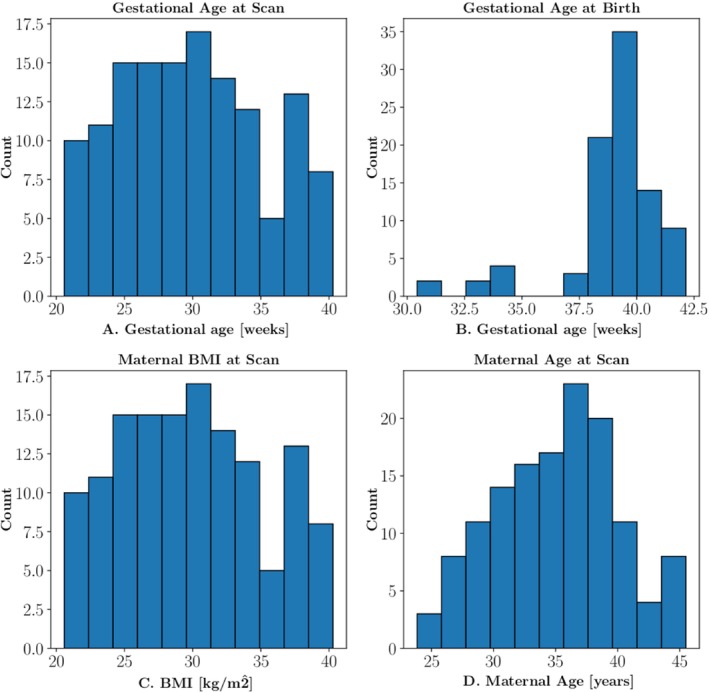
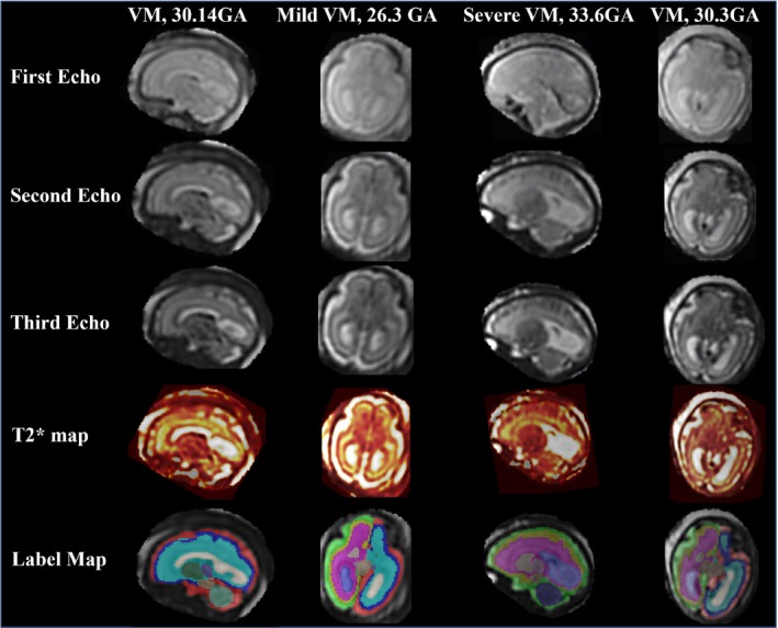
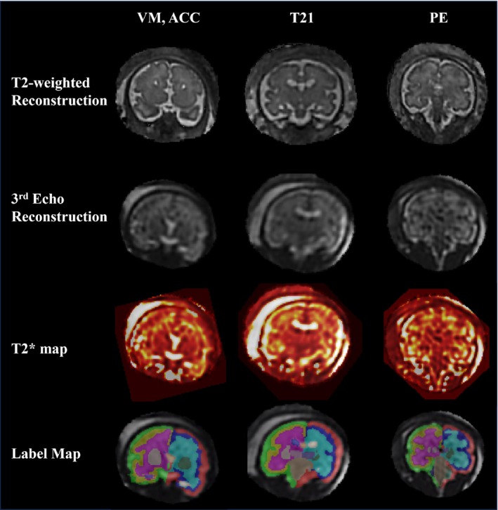
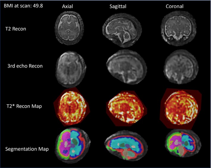
Dynamic multi-echo gradient-echo sequences were acquired in 135 cases at 0.55 T between 20 and 40 weeks' gestation. Automatic high-resolution reconstruction and segmentation tools were developed, resulting in T* values of seven individual anatomical brain structures for each subject. These regional brain T* values were analyzed throughout gestation.
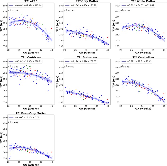
All regional fetal brain T* values decreased throughout gestation (p < 0.01). Each anatomical brain structure had varying ranges and decay rates, with the cerebellum and white matter displaying the highest (nonfluid structure) values, with the maximum values between 350 and 400 ms at about 20 weeks. The brainstem and deep gray matter had the lowest range of T* values, reaching values of 250 ms early in gestation. The matched volumetric assessment of the different structures demonstrated expected growth, matching current literature.
Low-field MRI allows for a detailed, regional T* analysis of the fetal brain, with more inclusive norms to be developed due to its wider bore.
妊娠期人类大脑发育复杂,因为结构和功能都在快速形成。利用磁共振成像(MRI)的结构成像方法已得到充分发展以探索这些变化,但功能成像工具却很匮乏。低场MRI是弥补这一差距的一种有前景的模式。低场强下较长的固有T值增加了动态范围,并能够对具有低T值的个体脑区进行量化,比如深部灰质。本研究在0.55T低场MRI上对整个妊娠后半期的脑区T*进行量化研究。
在妊娠20至40周期间,对135例孕妇进行了0.55T的动态多回波梯度回波序列扫描。开发了自动高分辨率重建和分割工具,得出了每个受试者7个个体解剖脑结构的T值。对整个妊娠期的这些脑区T值进行分析。
整个妊娠期所有胎儿脑区T值均下降(p<0.01)。每个解剖脑结构的范围和衰减率各不相同,小脑和白质显示出最高(非液体结构)值,在约20周时最大值在350至400毫秒之间。脑干和深部灰质的T值范围最低,在妊娠早期达到250毫秒的值。对不同结构的匹配体积评估显示出预期的生长情况,与当前文献相符。
低场MRI能够对胎儿脑进行详细的区域T*分析,由于其孔径更大,将制定更具包容性的标准。