Department of Pain Management, Shandong Provincial Hospital, Cheeloo College of Medicine, Shandong University, Jinan, Shandong 250021, P.R. China.
Department of Anesthesiology, Shandong Provincial Hospital Affiliated to Shandong First Medical University, Jinan, Shandong 250021, P.R. China.
Mol Med Rep. 2021 Jun;23(6). doi: 10.3892/mmr.2021.12067. Epub 2021 Apr 13.
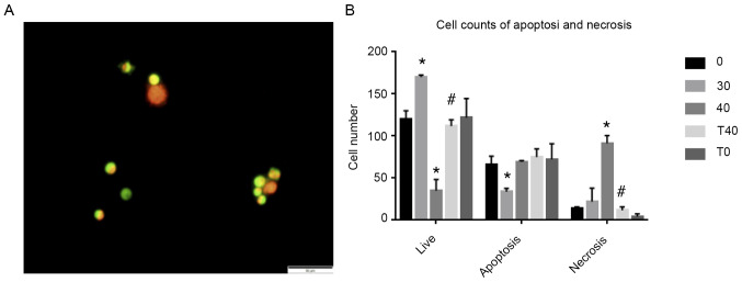
Ozone therapy can relieve multiple types of pain but exhibits potential neurotoxicity, the mechanism of which is unclear. The present study aimed to identify the role of nuclear factor (erythroid‑derived‑2)‑related 2 (NRF2) in preventing spinal cord injury caused by ozone overdose. Primary neuronal cells were extracted from newborn Wistar rats and authenticated by immunofluorescence using anti‑microtubule‑associated protein 2 as a cell type‑specific marker. Cell viability assay with different ozone concentrations (0, 10, 20, 30 and 40 g/ml) was used to determine the concentration that caused primary neuron injury; 30 min of 40 g/ml ozone therapy notably decreased cell viability to 71%. In order to test the effects of ozone, the cells were divided into five treatment groups [0‑, 30‑ and 40 g/ml ozone, tert‑butylhydroquinone (tBHQ) + 40 g/ml ozone (T40) and tBHQ (T0)]. Cells in the T40 and T0 groups received 40 mol/l tBHQ on the fifth day of SCN cultivation. Reverse transcription‑quantitative PCR and western blotting showed that protein expression levels of heme oxygenase‑1 (HO‑1) and mRNA expression levels of HO‑1 and NRF2 were decreased. NRF2, ubiquitin‑binding protein p62 and microtubule‑associated proteins 1A/1B light chain 3B expression levels were decreased following treatment with 40 g/ml ozone. Immunofluorescence showed that NRF2 nuclear expression levels also decreased following 40 g/ml ozone treatment. However, cells in the T40 group did not display decreased NRF2 nuclear expression levels. Normal/Apoptotic/Necrotic Cell Detection kit revealed that necrosis rate increased following treatment with 40 g/ml ozone; however, the T40 group did not exhibit this increased necrosis. At 40 g/ml, ozone increased spinal cord neuron (SCN) death . Moreover, treatment with 40 g/ml ozone damaged SCNs. The p62/NRF2/antioxidant response element pathway prevented such injury. tBHQ activated this pathway, upregulated autophagy and increased local nuclear NRF2 concentration, thus enhancing the antioxidant system to protect SCNs from injury caused by high concentrations of ozone.
臭氧疗法可缓解多种类型的疼痛,但具有潜在的神经毒性,其机制尚不清楚。本研究旨在确定核因子(红细胞衍生 2)相关 2(NRF2)在预防臭氧过量引起的脊髓损伤中的作用。从新生 Wistar 大鼠中提取原代神经元细胞,并通过抗微管相关蛋白 2 作为细胞类型特异性标志物进行免疫荧光鉴定。使用不同浓度(0、10、20、30 和 40 g/ml)的臭氧进行细胞活力测定,以确定引起原代神经元损伤的浓度;30 min 40 g/ml 臭氧治疗显著降低细胞活力至 71%。为了测试臭氧的作用,将细胞分为五组处理[0-、30-和 40 g/ml 臭氧、叔丁基对苯二酚(tBHQ)+40 g/ml 臭氧(T40)和 tBHQ(T0)]。在 SCN 培养的第 5 天,T40 和 T0 组的细胞接受 40 mol/l tBHQ。逆转录-定量 PCR 和 Western blot 显示血红素加氧酶 1(HO-1)蛋白表达水平和 HO-1 和 NRF2 的 mRNA 表达水平降低。40 g/ml 臭氧处理后 NRF2、泛素结合蛋白 p62 和微管相关蛋白 1A/1B 轻链 3B 的表达水平降低。免疫荧光显示 40 g/ml 臭氧处理后 NRF2 核表达水平也降低。然而,T40 组细胞中 NRF2 核表达水平并未降低。正常/凋亡/坏死细胞检测试剂盒显示,40 g/ml 臭氧处理后坏死率增加;然而,T40 组未显示出这种增加的坏死。在 40 g/ml 时,臭氧增加了脊髓神经元(SCN)的死亡。此外,40 g/ml 臭氧处理损伤 SCN。p62/NRF2/抗氧化反应元件通路防止了这种损伤。tBHQ 激活该通路,上调自噬并增加局部核 NRF2 浓度,从而增强抗氧化系统,保护 SCN 免受高浓度臭氧的损伤。