Imanishi Yuka, Hata Masaki, Matsukawa Ryohei, Aoyagi Atsushi, Omi Maiko, Mizutani Makoto, Naruse Keiko, Ozawa Shogo, Honda Masaki, Matsubara Tatsuaki, Takebe Jun
Department of Removable Prosthodontics, School of Dentistry, Aichi Gakuin University, 2-11 Suemori-dori, Chikusa-ku, Nagoya, 464-8651, Japan.
Department of Oral Anatomy, School of Dentistry, Aichi Gakuin University, Nagoya, Japan.
Inflamm Regen. 2021 Apr 14;41(1):12. doi: 10.1186/s41232-021-00163-w.
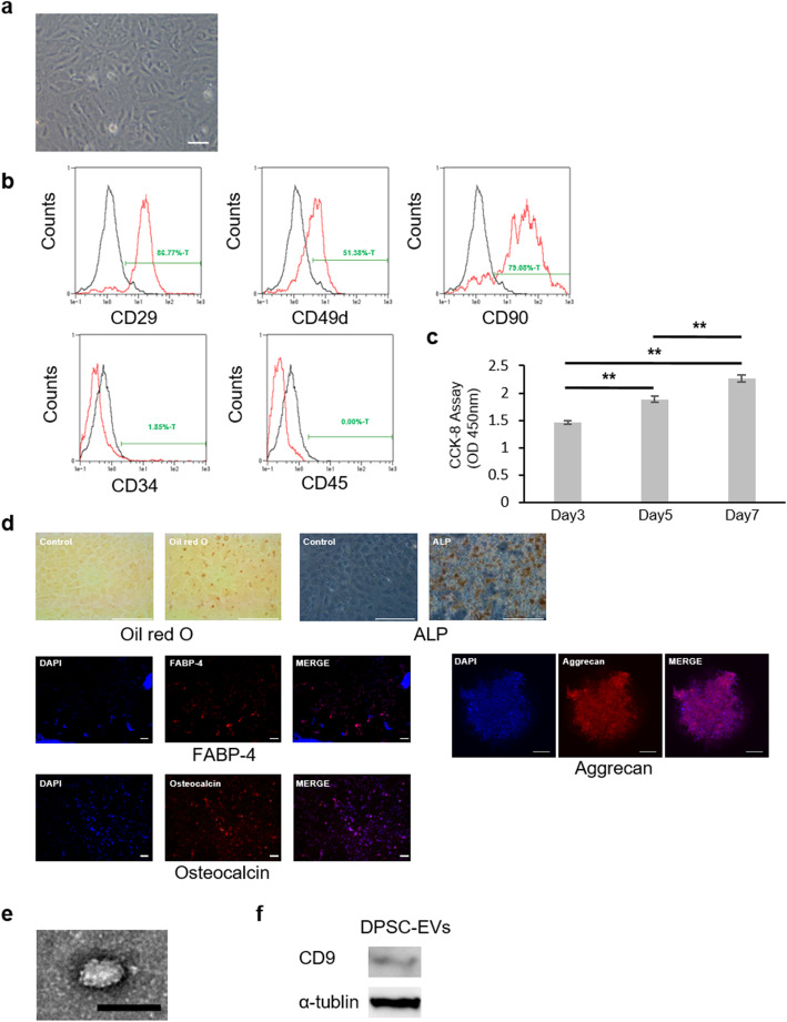
Extracellular vesicles (EVs) are known to be secreted by various cells. In particular, mesenchymal stem cell (MSC)-derived EVs (MSC-EVs) have tissue repair capacity and anti-inflammatory properties. Dental pulp stem cells (DPSCs), which are MSCs isolated from pulp tissue, are less invasive to the body than other MSCs and can be collected from young individuals. In this study, we investigated the efficacy of EVs secreted by DPSCs (DPSC-EVs) for bone formation.
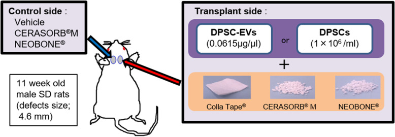
DPSC-EVs were isolated from the cell culture medium of DPSCs. DPSC-EVs were unilaterally injected along with collagen (COL), beta-tricalcium phosphate (β-TCP) or hydroxyapatite (HA) into rat calvarial bone defects. The effects of DPSC-EVs were analyzed by micro-computed tomography (micro-CT) and histological observation.
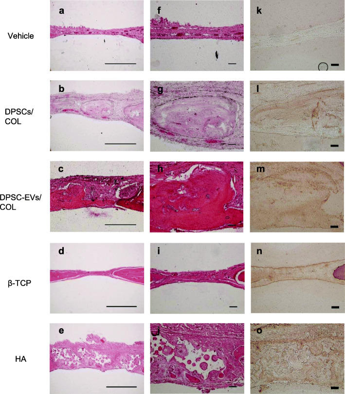
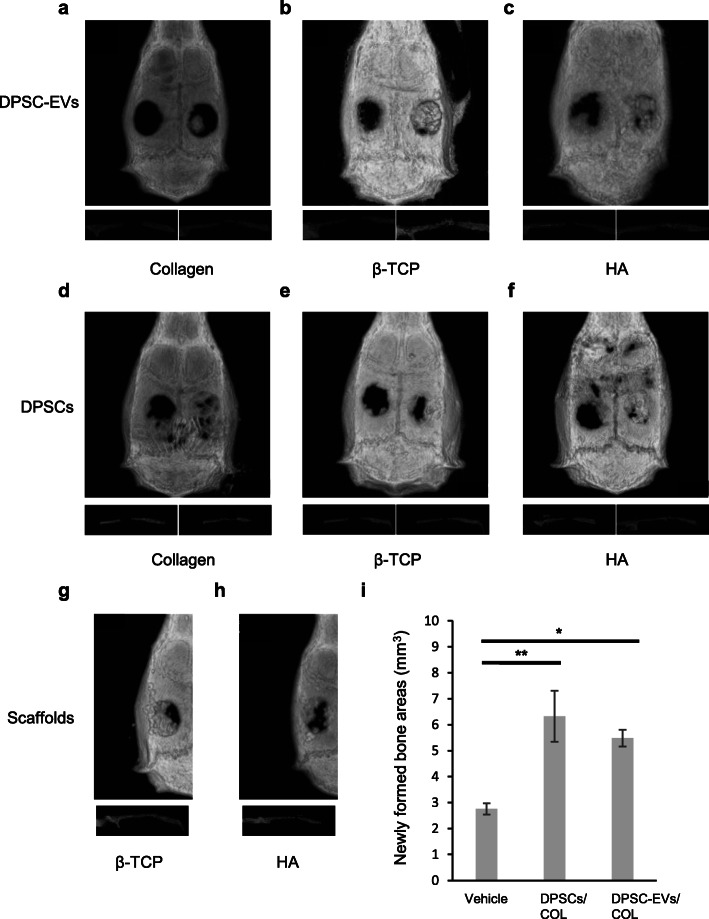
Micro-CT showed that administration of DPSC-EVs with the abovementioned scaffolds resulted in bone formation in the periphery of the defects. DPSC-EVs/COL specifically resulted in bone formation in the center of the defects. Histological observation revealed that DPSC-EVs/COL promoted new bone formation. Administration of DPSC-EVs/COL had almost the same effect on the bone defect site as transplantation of DPSCs/COL.
These results suggest that DPSC-EVs may be effective tools for bone tissue regeneration.
已知细胞外囊泡(EVs)由多种细胞分泌。特别是,间充质干细胞(MSC)衍生的EVs(MSC-EVs)具有组织修复能力和抗炎特性。牙髓干细胞(DPSCs)是从牙髓组织中分离出的间充质干细胞,对身体的侵入性比其他间充质干细胞小,并且可以从年轻个体中采集。在本研究中,我们研究了DPSCs分泌的EVs(DPSC-EVs)对骨形成的功效。
从DPSCs的细胞培养基中分离出DPSC-EVs。将DPSC-EVs与胶原蛋白(COL)、β-磷酸三钙(β-TCP)或羟基磷灰石(HA)一起单侧注射到大鼠颅骨骨缺损处。通过微计算机断层扫描(micro-CT)和组织学观察分析DPSC-EVs的作用。
Micro-CT显示,将DPSC-EVs与上述支架一起给药可导致缺损周边骨形成。DPSC-EVs/COL特别导致缺损中心骨形成。组织学观察显示DPSC-EVs/COL促进新骨形成。给予DPSC-EVs/COL对骨缺损部位的作用与移植DPSCs/COL几乎相同。
这些结果表明DPSC-EVs可能是骨组织再生的有效工具。