Coulson David J, Bakhashab Sherin, Latief Jevi Septyani, Weaver Jolanta U
Translational & Clinical Research Institute, Newcastle University, Newcastle Upon Tyne, NE2 4HH, UK.
Biochemistry Department, Faculty of Science, King Abdulaziz University, P.O. Box 80218, Jeddah, Saudi Arabia.
J Transl Med. 2021 Apr 16;19(1):140. doi: 10.1186/s12967-021-02785-7.
Type 1 diabetes (T1DM) is associated with premature cardiovascular disease (CVD) and a pro-inflammatory state whilst the proangiogenic miR-126-3p/-5p may play a role in CVD. Animal studies established miR-126 to be pro-angiogenic. We hypothesised miR-126-3p/-5p are reduced in T1DM whilst pro-inflammatory cytokines are increased.
29 well controlled, T1DM patients without CVD and 20 healthy controls (HCs) were studied. MiR-126-3p/-5p were assayed in plasma and peripheral blood mononuclear cells (PBMCs) whilst Chemokine C-X-C Receptor 1/2 (CXCR1/2) mRNA in PBMCs by real-time quantitative PCR. Cytokines were assayed by the Mesoscale Discovery. Ingenuity Pathway Analysis (IPA) was used to predict target genes, cellular functions and pathological states regulated by miR-126-3p/-5p. IPA generated both direct and indirect causations between different targets and analysed whether these effects would be inhibitory or stimulatory based on the published evidence.
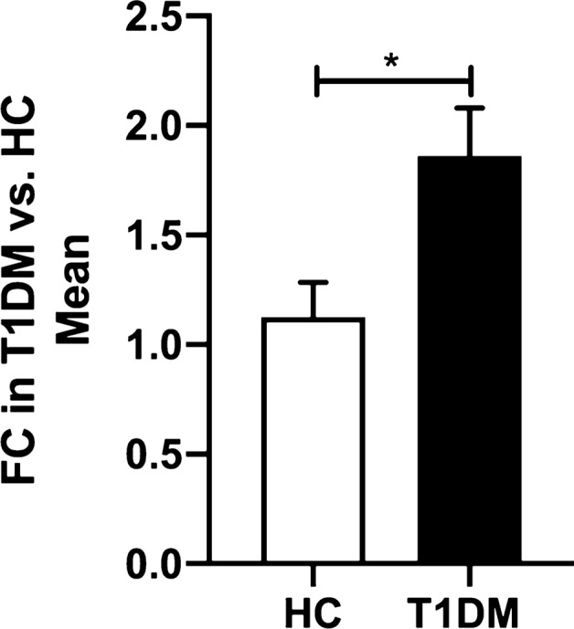
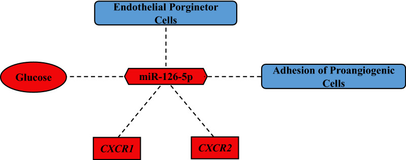
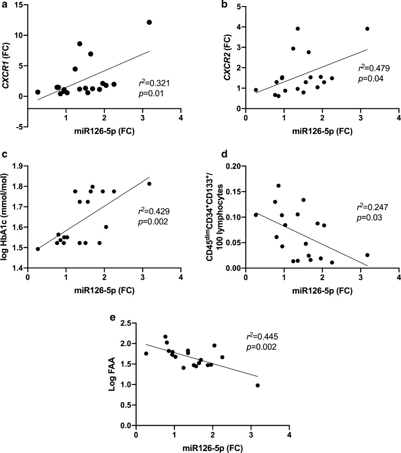
T1DM patients had a relatively good diabetic control (HbA1c = 7.4 ± 0.7% or 57.3 ± 7.6 mmol/mol). Homeostatic cytokine IL-7, pro-inflammatory cytokines IL-8 and TNF-α, and vascular endothelial growth factor-C (VEGF-C) were increased in T1DM, versus HCs; p = 0.008, p = 0.003, p = 0.041 and p = 0.013 respectively. MiR-126-5p was significantly upregulated in PBMCs in T1DM versus HCs; p = 0.01, but not in plasma. MiR-126-3p was unchanged. CXCR1/2 were elevated in T1DM versus HCs; p = 0.009 and p < 0.001 respectively. MiR-126-5p was positively correlated with CXCR1/2, and with HbA1c whilst negatively correlated with circulating endothelial progenitor cells (CD34CD133CD45) and fibronectin adhesion assay in a combined group of T1DM patients and HCs; p = 0.028 p = 0.049 p = 0.035 p = 0.047 and p = 0.004 respectively. IPA predicted miR-126-5p to be anti-inflammatory through the inhibition of chemokine C-C motif ligand 27, chymotrypsin-like elastase 2A and IL-7, whilst miR-126-3p had no direct anti-inflammatory effect. Simultaneously IPA predicted IL-7 as the most upstream cytokine target.
T1DM without apparent CVD or diabetic complications is an inflammatory state characterised not only by raised pro-inflammatory cytokines but also by increased receptor CXCR1/2 and miR-126-5p. MiR-126-5p upregulation may represent a compensatory response. Pro-miR-126-5p therapies or anti-IL-7 therapies may be a new option to reduce both inflammation and CVD risk in T1DM. Further research is required in a large prospective study in patients with T1DM.
1型糖尿病(T1DM)与心血管疾病(CVD)过早发生及促炎状态相关,而促血管生成的miR-126-3p/-5p可能在CVD中发挥作用。动物研究证实miR-126具有促血管生成作用。我们推测T1DM患者体内miR-126-3p/-5p水平降低,而促炎细胞因子水平升高。
对29例病情控制良好、无CVD的T1DM患者和20例健康对照者(HCs)进行研究。采用实时定量PCR检测血浆和外周血单个核细胞(PBMCs)中的miR-126-3p/-5p,同时检测PBMCs中趋化因子C-X-C受体1/2(CXCR1/2)的mRNA水平。采用中尺度发现法检测细胞因子。运用 Ingenuity 通路分析(IPA)预测miR-126-3p/-5p调控的靶基因、细胞功能和病理状态。IPA生成不同靶点之间的直接和间接因果关系,并根据已发表的证据分析这些效应是抑制性还是刺激性的。
T1DM患者的糖尿病控制情况相对良好(糖化血红蛋白A1c = 7.4 ± 0.7%或57.3 ± 7.6 mmol/mol)。与HCs相比,T1DM患者体内稳态细胞因子白细胞介素-7(IL-7)、促炎细胞因子白细胞介素-8(IL-8)和肿瘤坏死因子-α(TNF-α)以及血管内皮生长因子-C(VEGF-C)水平升高;p值分别为0.008、0.003、0.041和0.013。与HCs相比,T1DM患者PBMCs中的miR-126-5p显著上调;p = 0.01,但血浆中未出现此现象。miR-126-3p水平无变化。与HCs相比,T1DM患者的CXCR1/2水平升高;p值分别为0.009和p < 0.001。在T1DM患者和HCs的合并组中,miR-126-5p与CXCR1/2、糖化血红蛋白A1c呈正相关,而与循环内皮祖细胞(CD34CD133CD45)和纤连蛋白黏附试验呈负相关;p值分别为0.028、0.049、0.035、0.047和0.004。IPA预测miR-126-5p通过抑制趋化因子C-C基序配体27、类胰凝乳蛋白酶样弹性蛋白酶2A和IL-7发挥抗炎作用,而miR-126-3p无直接抗炎作用。同时,IPA预测IL-7是最上游的细胞因子靶点。
无明显CVD或糖尿病并发症的T1DM是一种炎症状态,其特征不仅在于促炎细胞因子升高,还在于受体CXCR1/2和miR-126-5p增加。miR-126-5p上调可能代表一种代偿反应。前体miR-126-5p疗法或抗IL-7疗法可能是降低T1DM炎症和CVD风险的新选择。需要对T1DM患者进行大规模前瞻性研究以作进一步探究。