Department of Ophthalmology, University of Oklahoma Health Sciences Center, Oklahoma City, OK, USA.
Department of Microbiology and Immunology, University of Oklahoma Health Sciences Center, Oklahoma City, OK, USA.
Exp Eye Res. 2021 Jun;207:108581. doi: 10.1016/j.exer.2021.108581. Epub 2021 Apr 15.
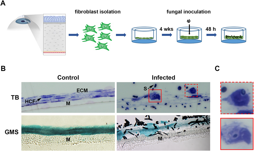
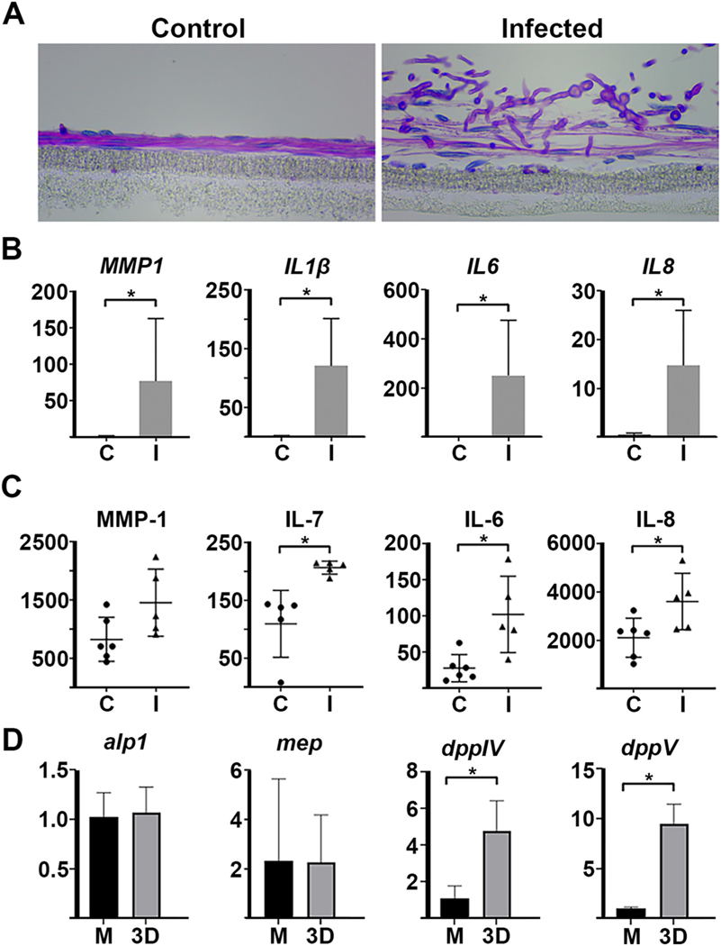
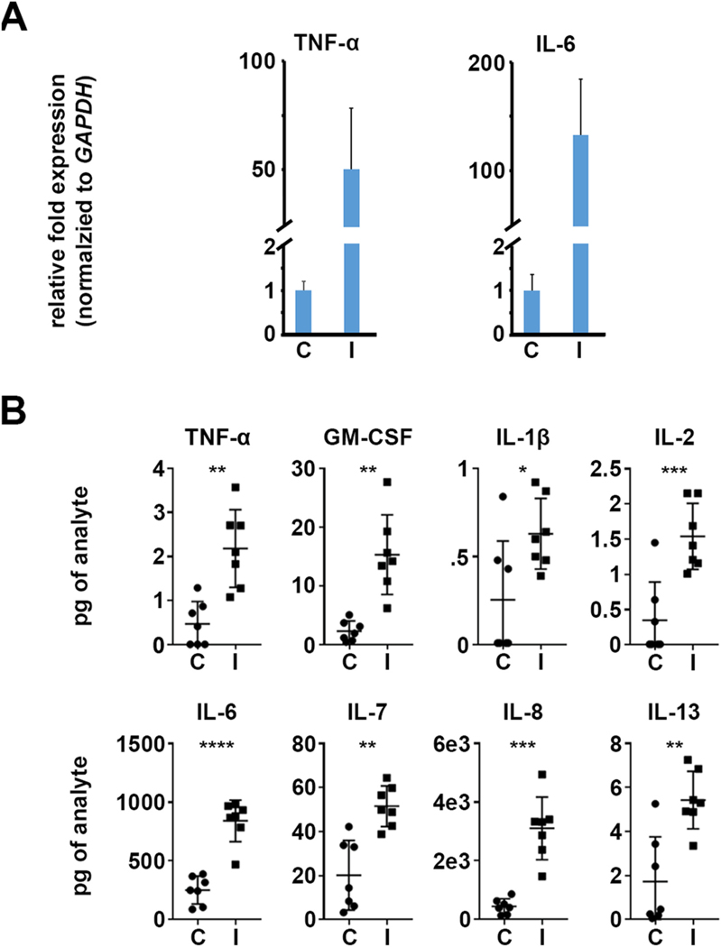
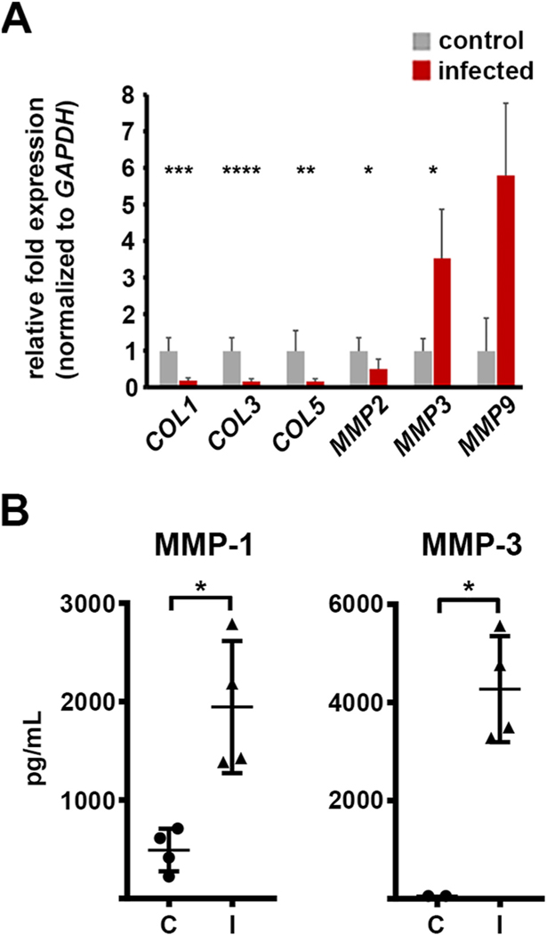
Fungal keratitis (FK) pathology is driven by both fungal growth and inflammation within the corneal stroma. Standard in vitro infection models ̶ involving co-culture of the pathogen and the corneal cells in tissue culture medium ̶ are sufficient to probe host responses to the fungus; however, they lack the physiological structure and nutrient composition of the stroma to accurately study fungal invasiveness and metabolic processes. We therefore sought to develop a culture model of FK that would allow for both host and fungal cell biology to be evaluated in parallel. Towards this end, we employed a previously described system in which primary human cornea fibroblasts (HCFs) are cultured on transwell membranes, whereupon they secrete a three-dimensional (3D) collagen matrix that resembles the human stroma. We demonstrated that two common mold agents of FK, Fusarium petroliphilum and Aspergillus fumigatus, penetrated into these constructs and caused a disruption of the collagen matrix that is characteristic of infection. HCF morphology appeared altered in the presence of fungus and electron microscopy revealed a clear internalization of fungal spores into these cells. Consistent with this apparent phagocyte-like activity of the HCFs, mRNA and protein levels for several pro-inflammatory cytokines/chemokines (including TNFα, IL-1β, IL-6, and IL-8) were significantly upregulated compared to uninfected samples. We similarly found an upregulation of several HCF metalloproteases (MMPs), which are enzymes that breakdown collagen during wound healing and may further activate pro-inflammatory signaling molecules. Finally, several fungal collagenase genes were upregulated during growth in the constructs relative to growth in tissue culture media alone, suggesting a fungal metabolic shift towards protein catabolism. Taken together, our results indicate that this 3D-stromal model provides a physiologically relevant system to study host and fungal cell pathobiology during FK.
真菌性角膜炎 (FK) 的发病机制是由真菌在角膜基质内的生长和炎症共同驱动的。标准的体外感染模型——涉及病原体与角膜细胞在组织培养基中的共培养——足以探测宿主对真菌的反应;然而,它们缺乏基质的生理结构和营养组成,无法准确研究真菌的侵袭性和代谢过程。因此,我们试图开发一种 FK 的培养模型,使宿主和真菌细胞生物学能够同时进行评估。为此,我们采用了先前描述的系统,其中原代人角膜成纤维细胞 (HCF) 培养在 Transwell 膜上,随后它们分泌出类似于人基质的三维 (3D) 胶原基质。我们证明,两种常见的 FK 霉菌剂,嗜油曲霉 (Fusarium petroliphilum) 和烟曲霉 (Aspergillus fumigatus),可以穿透这些结构,并导致胶原基质的破坏,这是感染的特征。在真菌存在的情况下,HCF 的形态似乎发生了改变,电子显微镜显示真菌孢子明显被内化到这些细胞中。与 HCF 明显的吞噬细胞样活性一致,几种促炎细胞因子/趋化因子(包括 TNFα、IL-1β、IL-6 和 IL-8)的 mRNA 和蛋白水平与未感染的样本相比显著上调。我们同样发现几种 HCF 金属蛋白酶 (MMPs) 的上调,这些酶在伤口愈合过程中分解胶原,可能进一步激活促炎信号分子。最后,与单独在组织培养基中生长相比,在这些结构中生长时,几种真菌胶原酶基因上调,表明真菌代谢向蛋白质分解代谢的转变。总之,我们的结果表明,这种 3D 基质模型为研究 FK 期间宿主和真菌细胞病理生物学提供了一个生理相关的系统。