Charité-Universitätsmedizin Berlin, corporate member of Freie Universität Berlin and Humboldt-Universität zu Berlin, Center for Stroke Research Berlin, Berlin, Germany.
Klinik und Hochschulambulanz für Neurologie, Charité - Universitätsmedizin Berlin, Berlin, Germany.
J Cereb Blood Flow Metab. 2021 Oct;41(10):2617-2627. doi: 10.1177/0271678X211010071. Epub 2021 Apr 17.
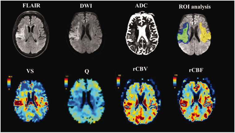
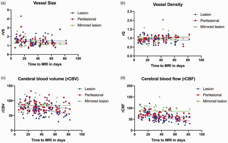
MRI-based vessel size imaging (VSI) allows for assessment of cerebral microvasculature and perfusion. This exploratory analysis of vessel size (VS) and density (Q; both assessed via VSI) in the subacute phase of ischemic stroke involved sixty-two patients from the BAPTISe cohort ('Biomarkers And Perfusion--Training-Induced changes after Stroke') nested within a randomized controlled trial (intervention: 4-week training relaxation). Relative VS, Q, cerebral blood volume (rCBV) and -flow (rCBF) were calculated for: ischemic lesion, perilesional tissue, and region corresponding to ischemic lesion on the contralateral side (mirrored lesion). Linear mixed-models detected significantly increased rVS and decreased rQ within the ischemic lesion compared to the mirrored lesion (coefficient[standard error]: 0.2[0.08] p = 0.03 and -1.0[0.3] p = 0.02, respectively); lesion rCBF and rCBV were also significantly reduced. Mixed-models did not identify time-to-MRI, nor training as modifying factors in terms of rVS or rQ up to two months post-stroke. Larger lesion VS was associated with larger lesion volumes (β 34, 95%CI 6.2-62; p = 0.02) and higher baseline NIHSS (β 3.0, 95%CI 0.49-5.3;p = 0.02), but was not predictive of six-month outcome. In summary, VSI can assess the cerebral microvasculature and tissue perfusion in the subacute phases of ischemic stroke, and may carry relevant prognostic value in terms of lesion volume and stroke severity.
MRI 血管大小成像(VSI)可用于评估脑微循环和灌注。这项对缺血性卒中亚急性期血管大小(VS)和密度(Q;均通过 VSI 评估)的探索性分析涉及 BAPTISe 队列('Biomarkers And Perfusion--Training-Induced changes after Stroke')中的 62 名患者,该队列嵌套在一项随机对照试验中(干预:4 周训练放松)。相对 VS、Q、脑血容量(rCBV)和血流(rCBF)在:缺血性病变、病变周围组织以及对侧与缺血性病变相对应的区域(镜像病变)中进行了计算。线性混合模型检测到与镜像病变相比,缺血性病变内 rVS 显著增加,rQ 显著降低(系数[标准误差]:0.2[0.08]p=0.03 和-1.0[0.3]p=0.02,分别);病变 rCBF 和 rCBV 也显著降低。混合模型未在两个月内发现 MRI 时间或训练对 rVS 或 rQ 的影响,也未发现时间或训练是其影响因素。较大的病变 VS 与较大的病变体积(β 34,95%CI 6.2-62;p=0.02)和较高的基线 NIHSS(β 3.0,95%CI 0.49-5.3;p=0.02)相关,但不能预测六个月的结果。总之,VSI 可评估缺血性卒中亚急性期的脑微血管和组织灌注,并且在病变体积和卒中严重程度方面可能具有相关的预后价值。