Dong Shuang-Shuang, Dong Dan-Dan, Yang Zhang-Fu, Zhu Gui-Qi, Gao Dong-Mei, Chen Jie, Zhao Yan, Liu Bin-Bin
Liver Cancer Institute, Zhongshan Hospital, Fudan University and Key Laboratory of Carcinogenesis and Cancer Invasion, Ministry of Education, Shanghai, China.
Department of Biochemistry and Molecular Biology, School of Basic Medical Sciences, Fudan University, Shanghai, China.
Front Cell Dev Biol. 2021 Mar 29;9:633358. doi: 10.3389/fcell.2021.633358. eCollection 2021.
Angiogenesis is a crucial process in tumorigenesis and development. The role of exosomes derived from hepatocellular carcinoma (HCC) cells in angiogenesis has not been clearly elucidated.
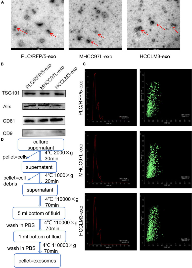
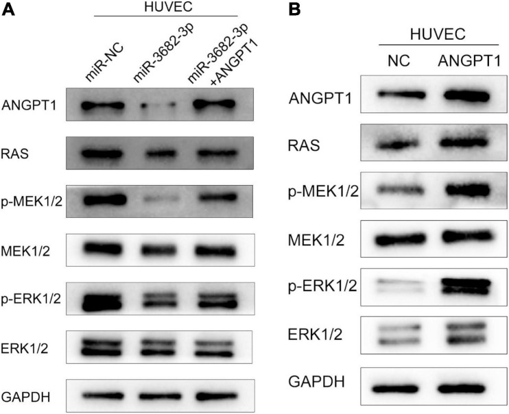
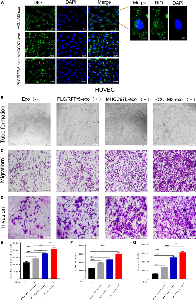
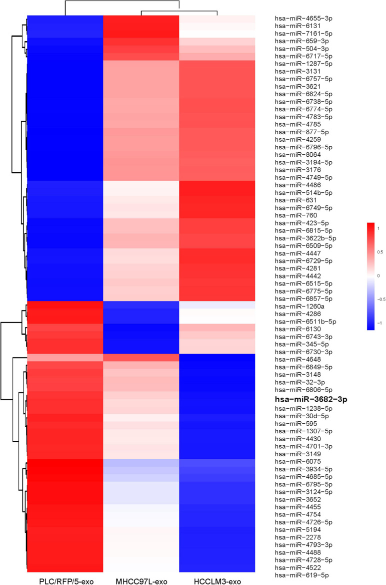
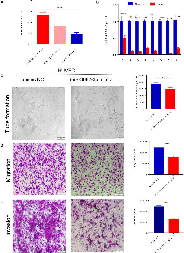
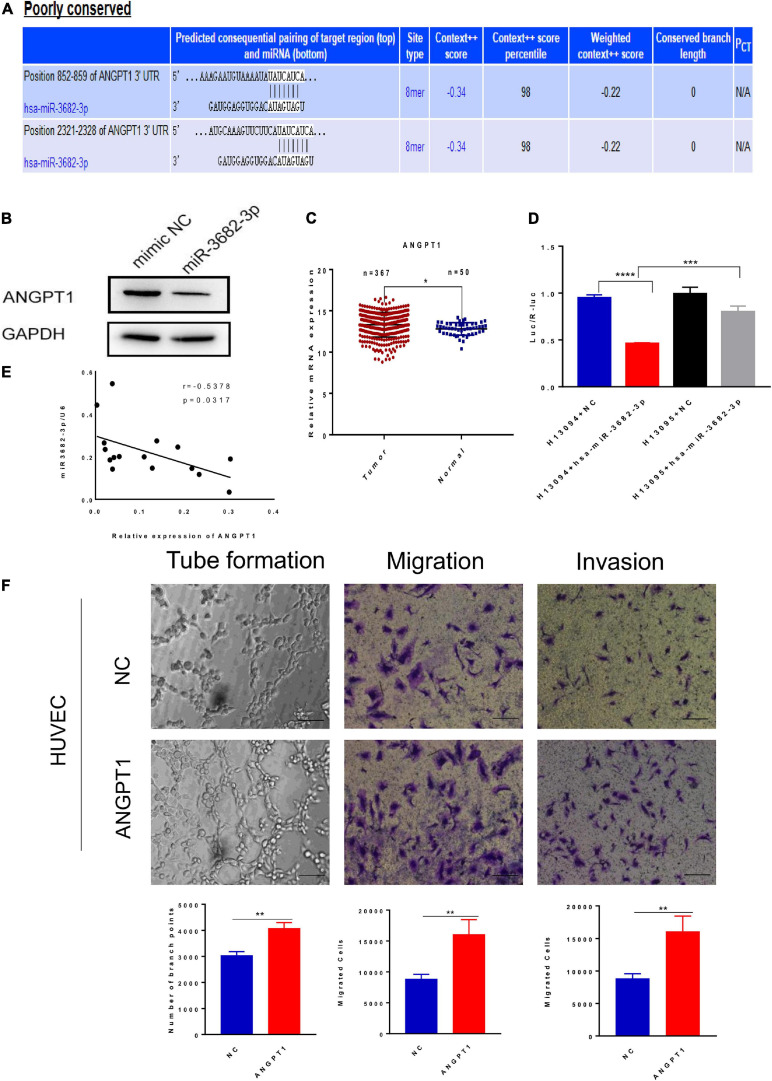
Exosomes were isolated from HCC cell lines (HCCLM3, MHCC97L, and PLC/RFP/5) by ultracentrifugation and identified by nano transmission electron microscopy (TEM), NanoSight analysis and western blotting, respectively. and analyses showed that exosomes isolated from highly metastatic HCC cells enhanced the migration, invasion and tube formation of human umbilical vein endothelial cells (HUVECs) compared to exosomes derived from poorly metastatic HCC cells. In addition, microarray analysis of HCC-Exos was conducted to identify potential functional molecules, and miR-3682-3p expression was found to be significantly downregulated in exosomes isolated from highly metastatic HCC cells. By gain-of-function experiments, we found that HCC cells secreted exosomal miR-3682-3p, which negatively regulates angiopoietin-1 (ANGPT1), and this led to inhibition of RAS-MEK1/2-ERK1/2 signaling in endothelial cells and eventually impaired angiogenesis.
Our study elucidates that exosomal miR-3682-3p attenuates angiogenesis by targeting ANGPT1 through RAS-MEK1/2-ERK1/2 signaling and provides novel potential targets for liver cancer therapy.
血管生成是肿瘤发生和发展中的关键过程。肝细胞癌(HCC)细胞来源的外泌体在血管生成中的作用尚未明确阐明。
通过超速离心从肝癌细胞系(HCCLM3、MHCC97L和PLC/RFP/5)中分离外泌体,并分别通过纳米透射电子显微镜(TEM)、纳米可视分析和蛋白质印迹法进行鉴定。 和 分析表明,与低转移肝癌细胞来源的外泌体相比,高转移肝癌细胞来源的外泌体增强了人脐静脉内皮细胞(HUVECs)的迁移、侵袭和管腔形成。此外,对肝癌外泌体进行微阵列分析以鉴定潜在的功能分子,发现高转移肝癌细胞来源的外泌体中miR-3682-3p表达显著下调。通过功能获得实验,我们发现肝癌细胞分泌外泌体miR-3682-3p,其负向调节血管生成素-1(ANGPT1),并导致内皮细胞中RAS-MEK1/2-ERK1/2信号传导受到抑制,最终损害血管生成。
我们的研究阐明外泌体miR-3682-3p通过RAS-MEK1/2-ERK1/2信号传导靶向ANGPT1来减弱血管生成,并为肝癌治疗提供了新的潜在靶点。