Buffalo Neuroimaging Analysis Center, Department of Neurology, School of Medicine and Biomedical Sciences, University at Buffalo, State University of New York, Buffalo, NY, USA; Jacobs MS Center, Department of Neurology, Jacobs School of Medicine and Biomedical Sciences, University at Buffalo, State University of New York, Buffalo, NY, USA.
Buffalo Neuroimaging Analysis Center, Department of Neurology, School of Medicine and Biomedical Sciences, University at Buffalo, State University of New York, Buffalo, NY, USA.
Neuroimage Clin. 2021;30:102652. doi: 10.1016/j.nicl.2021.102652. Epub 2021 Mar 29.
Thalamic volume loss is a key marker of neurodegeneration in multiple sclerosis (MS). T2-FLAIR MRI is a common denominator in clinical routine MS imaging, but current methods for thalamic volumetry are not applicable to it.
To develop and validate a robust algorithm to measure thalamic volume using clinical routine T2-FLAIR MRI.
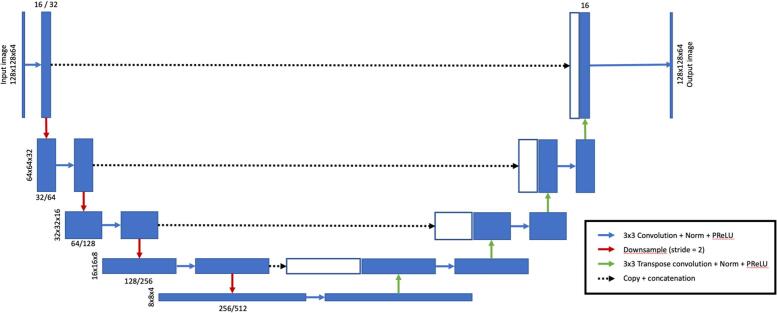
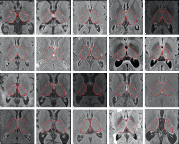
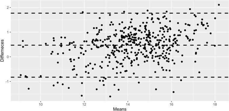
A dual-stage deep learning approach based on 3D U-net (DeepGRAI - Deep Gray Rating via Artificial Intelligence) was created and trained/validated/tested on 4,590 MRI exams (4288 2D-FLAIR, 302 3D-FLAIR) from 59 centers (80/10/10 train/validation/test split). As training/test targets, FIRST was used to generate thalamic masks from 3D T1 images. Masks were reviewed, corrected, and aligned into T2-FLAIR space. Additional validation was performed to assess inter-scanner reliability (177 subjects at 1.5 T and 3 T within one week) and scan-rescan-reliability (5 subjects scanned, repositioned, and then re-scanned). A longitudinal dataset including assessment of disability and cognition was used to evaluate the predictive value of the approach.
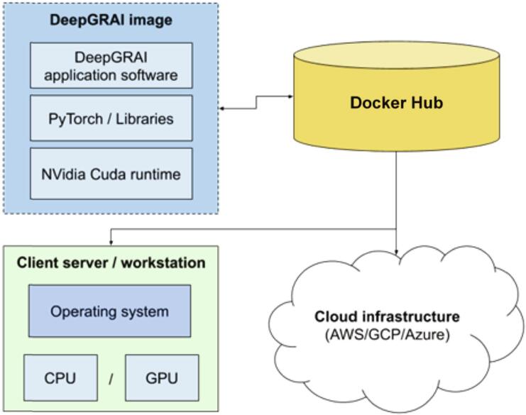
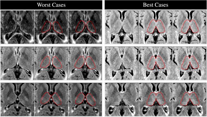
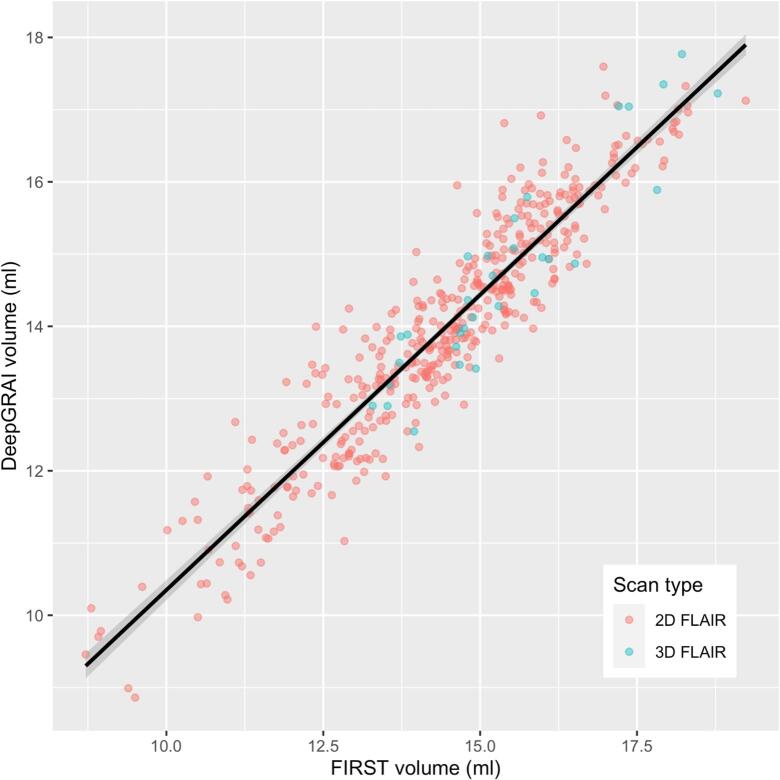
DeepGRAI automatically quantified thalamic volume in approximately 7 s per case, and has been made publicly available. Accuracy on T2-FLAIR relative to 3D T1 FIRST was 99.4% (r = 0.94, p < 0.001,TPR = 93.0%, FPR = 0.3%). Inter-scanner error was 3.21%. Scan-rescan error with repositioning was 0.43%. DeepGRAI-derived thalamic volume was associated with disability (r = -0.427,p < 0.001) and cognition (r = -0.537,p < 0.001), and was a significant predictor of longitudinal cognitive decline (R = 0.081, p = 0.024; comparatively, FIRST-derived volume was R = 0.080, p = 0.025).
DeepGRAI provides fast, reliable, and clinically relevant thalamic volume measurement on multicenter clinical-quality T2-FLAIR images. This indicates potential for real-world thalamic volumetry, as well as quantification on legacy datasets without 3D T1 imaging.
丘脑体积损失是多发性硬化症(MS)神经退行性变的一个关键标志物。T2-FLAIR MRI 是临床常规 MS 成像中的一个共同点,但目前用于丘脑体积测量的方法并不适用于 T2-FLAIR MRI。
开发和验证一种使用临床常规 T2-FLAIR MRI 测量丘脑体积的强大算法。
创建了一种基于 3D U-net(通过人工智能进行深灰度分级-Deep Gray Rating via Artificial Intelligence)的双阶段深度学习方法,并在来自 59 个中心的 4590 次 MRI 检查(4288 次 2D-FLAIR,302 次 3D-FLAIR)上进行了训练/验证/测试(80/10/10 训练/验证/测试分割)。作为训练/测试目标,使用 FIRST 从 3D T1 图像生成丘脑掩模。对掩模进行了审查、纠正,并对齐到 T2-FLAIR 空间。还进行了额外的验证,以评估扫描仪间的可靠性(在一周内对 1.5T 和 3T 中的 177 名受试者进行评估)和扫描-重扫可靠性(对 5 名受试者进行扫描、重新定位,然后再次扫描)。使用包括评估残疾和认知的纵向数据集来评估该方法的预测价值。
DeepGRAI 平均每例大约 7s 自动量化丘脑体积,并且已经公开。相对于 3D T1 FIRST 的 T2-FLAIR 准确性为 99.4%(r=0.94,p<0.001,TPR=93.0%,FPR=0.3%)。扫描仪间误差为 3.21%。重新定位的扫描-重扫误差为 0.43%。DeepGRAI 衍生的丘脑体积与残疾(r=-0.427,p<0.001)和认知(r=-0.537,p<0.001)相关,是纵向认知下降的显著预测因子(R=0.081,p=0.024;相比之下,FIRST 衍生的体积为 R=0.080,p=0.025)。
DeepGRAI 可在多中心临床质量 T2-FLAIR 图像上快速、可靠且具有临床相关性地提供丘脑体积测量。这表明该方法具有在没有 3D T1 成像的情况下进行实际的丘脑体积测量和量化的潜力。