• 文献检索
  • 文档翻译
  • 深度研究
  • 学术资讯
  • Suppr Zotero 插件Zotero 插件
  • 邀请有礼
  • 套餐&价格
  • 历史记录
应用&插件
Suppr Zotero 插件Zotero 插件浏览器插件Mac 客户端Windows 客户端微信小程序
定价
高级版会员购买积分包购买API积分包
服务
文献检索文档翻译深度研究API 文档MCP 服务
关于我们
关于 Suppr公司介绍联系我们用户协议隐私条款
关注我们

Suppr 超能文献

核心技术专利:CN118964589B侵权必究
粤ICP备2023148730 号-1Suppr @ 2026

文献检索

告别复杂PubMed语法,用中文像聊天一样搜索,搜遍4000万医学文献。AI智能推荐,让科研检索更轻松。

立即免费搜索

文件翻译

保留排版,准确专业,支持PDF/Word/PPT等文件格式,支持 12+语言互译。

免费翻译文档

深度研究

AI帮你快速写综述,25分钟生成高质量综述,智能提取关键信息,辅助科研写作。

立即免费体验

基于雅可比积分的病理性深部灰质和丘脑体积损失的年龄依赖性截断值。

Age-dependent cut-offs for pathological deep gray matter and thalamic volume loss using Jacobian integration.

机构信息

jung diagnostics GmbH, Hamburg, Germany.

jung diagnostics GmbH, Hamburg, Germany.

出版信息

Neuroimage Clin. 2020;28:102478. doi: 10.1016/j.nicl.2020.102478. Epub 2020 Oct 27.

DOI:10.1016/j.nicl.2020.102478
PMID:33269702
原文链接:https://pmc.ncbi.nlm.nih.gov/articles/PMC7645288/
Abstract

INTRODUCTION

Several recent studies indicate that deep gray matter or thalamic volume loss (VL) might be promising surrogate markers of disease activity in multiple sclerosis (MS) patients. To allow applying these markers to individual MS patients in clinical routine, age-dependent cut-offs distinguishing physiological from pathological VL and an estimation of the measurement error, which provides the confidence of the result, are to be defined.

METHODS

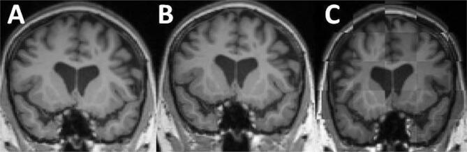
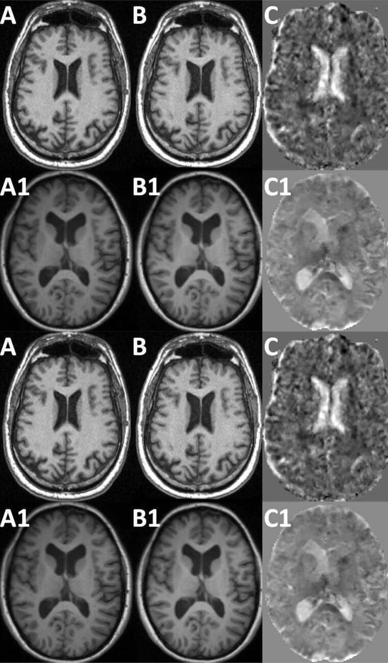
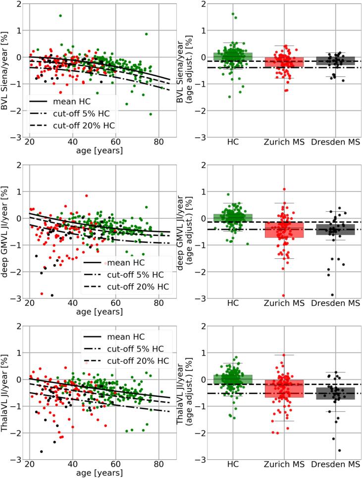
Longitudinal MRI scans of the following cohorts were analyzed in this study: 189 healthy controls (HC) (mean age 54 years, 22% female), 98 MS patients from Zurich university hospital (mean age 34 years, 62% female), 33 MS patients from Dresden university hospital (mean age 38 years, 60% female), and publicly available reliability data sets consisting of 162 short-term MRI scan-rescan pairs with scan intervals of days or few weeks. Percentage annualized whole brain volume loss (BVL), gray matter (GM) volume loss (GMVL), deep gray matter volume loss (deep GMVL), and thalamic volume loss (ThalaVL) were computed deploying the Jacobian integration (JI) method. BVL was additionally computed using Siena, an established method used in many Phase III drug trials. A linear mixed effect model was used to estimate the measurement error as the standard deviation (SD) of model residuals of all 162 scan-rescan pairs For estimation of age-dependent cut-offs, a quadratic regression function between age and the corresponding annualized VL values of the HC was computed. The 5th percentile was defined as the threshold for pathological VL per year since 95% of HC subjects exhibit a less pronounced VL for a given age. For the MS patients BVL, GMVL, deep GMVL, and ThalaVL were mutually compared and a paired t-test was used to test whether there are systematic differences in VL between these brain regions.

RESULTS

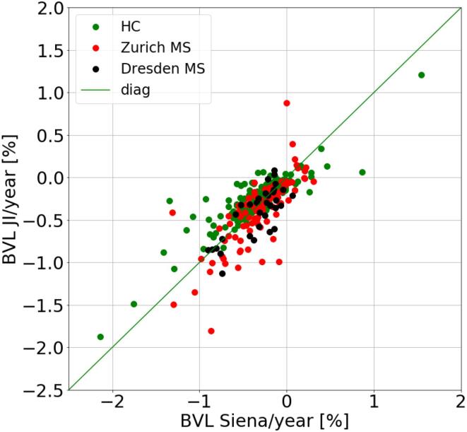
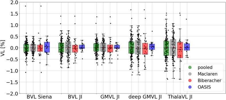
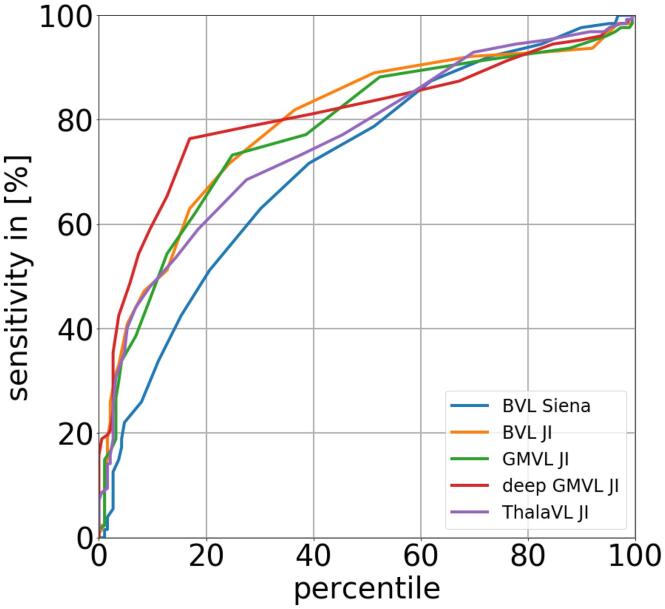
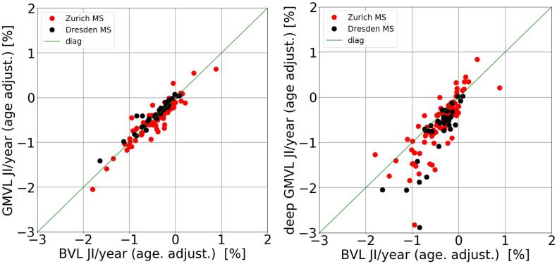
Siena and JI showed a high agreement for BVL measures, with a median absolute difference of 0.1% and a correlation coefficient of r = 0.78. Siena and GMVL showed a similar standard deviation (SD) of the scan-rescan error of 0.28% and 0.29%, respectively. For deep GMVL, ThalaVL the SD of the scan-rescan error was slightly higher (0.43% and 0.5%, respectively). Among the HC the thalamus showed the highest mean VL (-0.16%, -0.39%, and -0.59% at ages 35, 55, and 75, respectively). Corresponding cut-offs for a pathological VL/year were -0.68%, -0.91%, and -1.11%. The MS cohorts did not differ in BVL and GMVL. However, both MS cohorts showed a significantly (p = 0.05) stronger deep GMVL than BVL per year.

CONCLUSION

It might be methodologically feasible to assess deep GMVL using JI in individual MS patients. However, age and the measurement error need to be taken into account. Furthermore, deep GMVL may be used as a complementary marker to BVL since MS patients exhibit a significantly stronger deep GMVL than BVL.

摘要

简介

最近的几项研究表明,深部灰质或丘脑体积损失(VL)可能是多发性硬化症(MS)患者疾病活动的有前途的替代标志物。为了在临床常规中应用这些标志物于个体 MS 患者,需要定义区分生理性与病理性 VL 的年龄依赖性截止值,并评估测量误差,这提供了结果的置信度。

方法

本研究分析了以下队列的纵向 MRI 扫描:189 名健康对照者(HC)(平均年龄 54 岁,22%为女性)、98 名来自苏黎世大学医院的 MS 患者(平均年龄 34 岁,62%为女性)、33 名来自德累斯顿大学医院的 MS 患者(平均年龄 38 岁,60%为女性),以及由 162 个短期 MRI 扫描-再扫描对组成的公开可得的可靠性数据集,扫描间隔为几天或数周。使用 Jacobian 积分(JI)方法计算了每年的全脑体积损失(BVL)、灰质(GM)体积损失(GMVL)、深部灰质体积损失(深部 GMVL)和丘脑体积损失(ThalaVL)的百分比。BVL 还使用 Siena 进行了计算,Siena 是许多 III 期药物试验中使用的一种成熟方法。使用线性混合效应模型来估计测量误差,即所有 162 个扫描-再扫描对的模型残差的标准差(SD)。为了估计年龄依赖性截止值,计算了 HC 年龄与相应年度 VL 值之间的二次回归函数。第 5 个百分位数被定义为每年病理性 VL 的阈值,因为 95%的 HC 受试者在给定年龄时表现出较不明显的 VL。对于 MS 患者的 BVL、GMVL、深部 GMVL 和 ThalaVL,进行了相互比较,并使用配对 t 检验检验这些脑区之间的 VL 是否存在系统差异。

结果

Siena 和 JI 对 BVL 测量值具有高度一致性,中位数绝对差值为 0.1%,相关系数 r=0.78。Siena 和 GMVL 的扫描-再扫描误差的标准偏差(SD)分别为 0.28%和 0.29%。对于深部 GMVL 和 ThalaVL,扫描-再扫描误差的 SD 略高(分别为 0.43%和 0.5%)。在 HC 中,丘脑的平均 VL 最高(分别为 35、55 和 75 岁时的-0.16%、-0.39%和-0.59%)。相应的每年病理性 VL 截止值分别为-0.68%、-0.91%和-1.11%。MS 队列在 BVL 和 GMVL 方面没有差异。然而,两个 MS 队列的深部 GMVL 每年都明显(p=0.05)强于 BVL。

结论

在个体 MS 患者中使用 JI 评估深部 GMVL 在方法学上可能是可行的。然而,需要考虑年龄和测量误差。此外,深部 GMVL 可用作 BVL 的补充标志物,因为 MS 患者的深部 GMVL 明显强于 BVL。

https://cdn.ncbi.nlm.nih.gov/pmc/blobs/6d30/7645288/e99420709687/gr7.jpg
https://cdn.ncbi.nlm.nih.gov/pmc/blobs/6d30/7645288/64a52cfc8af3/gr1.jpg
https://cdn.ncbi.nlm.nih.gov/pmc/blobs/6d30/7645288/869a4ba4dbae/gr2.jpg
https://cdn.ncbi.nlm.nih.gov/pmc/blobs/6d30/7645288/545adb75a20e/gr3.jpg
https://cdn.ncbi.nlm.nih.gov/pmc/blobs/6d30/7645288/c885ac0a05de/gr4.jpg
https://cdn.ncbi.nlm.nih.gov/pmc/blobs/6d30/7645288/49ed1d4869a1/gr5.jpg
https://cdn.ncbi.nlm.nih.gov/pmc/blobs/6d30/7645288/05907ac6ec84/gr6.jpg
https://cdn.ncbi.nlm.nih.gov/pmc/blobs/6d30/7645288/e99420709687/gr7.jpg

相似文献

1
Age-dependent cut-offs for pathological deep gray matter and thalamic volume loss using Jacobian integration.基于雅可比积分的病理性深部灰质和丘脑体积损失的年龄依赖性截断值。
Neuroimage Clin. 2020;28:102478. doi: 10.1016/j.nicl.2020.102478. Epub 2020 Oct 27.
2
Assessing Biological and Methodological Aspects of Brain Volume Loss in Multiple Sclerosis.评估多发性硬化症中脑容量损失的生物学和方法学方面。
JAMA Neurol. 2018 Oct 1;75(10):1246-1255. doi: 10.1001/jamaneurol.2018.1596.
3
SIENA-XL for improving the assessment of gray and white matter volume changes on brain MRI.SIENA-XL 可改善脑 MRI 上灰质和白质体积变化的评估。
Hum Brain Mapp. 2018 Mar;39(3):1063-1077. doi: 10.1002/hbm.23828. Epub 2017 Dec 8.
4
Within-patient fluctuation of brain volume estimates from short-term repeated MRI measurements using SIENA/FSL.利用 SIENA/FSL 进行短期重复 MRI 测量时脑容量估计的个体内波动。
J Neurol. 2018 May;265(5):1158-1165. doi: 10.1007/s00415-018-8825-8. Epub 2018 Mar 16.
5
Global and regional annual brain volume loss rates in physiological aging.生理衰老过程中的全球及区域年度脑容量损失率。
J Neurol. 2017 Mar;264(3):520-528. doi: 10.1007/s00415-016-8374-y. Epub 2017 Jan 4.
6
Pathological cut-offs of global and regional brain volume loss in multiple sclerosis.多发性硬化症中全脑和区域性脑容量损失的病理性截止值。
Mult Scler. 2019 Apr;25(4):541-553. doi: 10.1177/1352458517742739. Epub 2017 Nov 16.
7
Estimates of age-dependent cutoffs for pathological brain volume loss using SIENA/FSL-a longitudinal brain volumetry study in healthy adults.使用 SIENA/FSL 对健康成年人进行的纵向脑容积研究估计病理性脑容积丢失的年龄依赖性截断值。
Neurobiol Aging. 2018 May;65:1-6. doi: 10.1016/j.neurobiolaging.2017.12.024. Epub 2017 Dec 30.
8
Determinants of Deep Gray Matter Atrophy in Multiple Sclerosis: A Multimodal MRI Study.多发性硬化症深部灰质萎缩的决定因素:一项多模态 MRI 研究。
AJNR Am J Neuroradiol. 2019 Jan;40(1):99-106. doi: 10.3174/ajnr.A5915. Epub 2018 Dec 20.
9
Association of volumetric MRI measures and disability in MS patients of the same age: Descriptions from a birth year cohort.磁共振容积成像测量与同龄 MS 患者残疾的相关性:来自同一年龄队列的描述。
Mult Scler Relat Disord. 2023 Mar;71:104568. doi: 10.1016/j.msard.2023.104568. Epub 2023 Feb 12.
10
Methods for Brain Atrophy MR Quantification in Multiple Sclerosis: Application to the Multicenter INNI Dataset.多发性硬化症脑萎缩磁共振定量分析方法:多中心 INNI 数据集的应用。
J Magn Reson Imaging. 2023 Oct;58(4):1221-1231. doi: 10.1002/jmri.28616. Epub 2023 Jan 20.

引用本文的文献

1
Patients with relapsing-remitting multiple sclerosis show accelerated whole brain volume and thalamic volume loss early in disease.复发缓解型多发性硬化症患者在疾病早期会出现全脑体积和丘脑体积加速丢失的情况。
Neuroradiology. 2025 Jan;67(1):99-107. doi: 10.1007/s00234-024-03516-7. Epub 2024 Nov 28.
2
BrainLossNet: a fast, accurate and robust method to estimate brain volume loss from longitudinal MRI.BrainLossNet:一种快速、准确且稳健的方法,可从纵向 MRI 估计脑容量损失。
Int J Comput Assist Radiol Surg. 2024 Sep;19(9):1763-1771. doi: 10.1007/s11548-024-03201-3. Epub 2024 Jun 16.
3
Ocrelizumab-treated patients with relapsing multiple sclerosis show volume loss rates similar to healthy aging.

本文引用的文献

1
Fully automated longitudinal segmentation of new or enlarged multiple sclerosis lesions using 3D convolutional neural networks.使用 3D 卷积神经网络全自动纵向分割新的或扩大的多发性硬化病变。
Neuroimage Clin. 2020;28:102445. doi: 10.1016/j.nicl.2020.102445. Epub 2020 Sep 24.
2
Contribution of normal aging to brain atrophy in MS.正常衰老对 MS 脑萎缩的影响。
Neurol Neuroimmunol Neuroinflamm. 2019 Sep 25;6(6). doi: 10.1212/NXI.0000000000000616. Print 2019 Nov.
3
Brain volume loss in individuals over time: Source of variance and limits of detectability.
奥瑞珠单抗治疗的复发型多发性硬化症患者的容积损失率与健康衰老相似。
Mult Scler. 2023 May;29(6):741-747. doi: 10.1177/13524585231162586. Epub 2023 May 6.
4
Commercial volumetric MRI reporting tools in multiple sclerosis: a systematic review of the evidence.商业容积磁共振成像报告工具在多发性硬化症中的应用:系统评价证据。
Neuroradiology. 2023 Jan;65(1):5-24. doi: 10.1007/s00234-022-03074-w. Epub 2022 Nov 4.
5
Single-subject analysis of regional brain volumetric measures can be strongly influenced by the method for head size adjustment.对区域性脑容量测量的单个体分析可能会受到头部大小调整方法的强烈影响。
Neuroradiology. 2022 Oct;64(10):2001-2009. doi: 10.1007/s00234-022-02961-6. Epub 2022 Apr 25.
个体脑容量随时间的损失:方差来源与可检测性的局限性。
Neuroimage. 2020 Jul 1;214:116737. doi: 10.1016/j.neuroimage.2020.116737. Epub 2020 Mar 21.
4
Safety and efficacy of ozanimod versus interferon beta-1a in relapsing multiple sclerosis (RADIANCE): a multicentre, randomised, 24-month, phase 3 trial.奥扎莫德与干扰素β-1a 在复发型多发性硬化症(RADIANCE)中的安全性和疗效:一项多中心、随机、24 个月、3 期临床试验。
Lancet Neurol. 2019 Nov;18(11):1021-1033. doi: 10.1016/S1474-4422(19)30238-8. Epub 2019 Sep 3.
5
Safety and efficacy of ozanimod versus interferon beta-1a in relapsing multiple sclerosis (SUNBEAM): a multicentre, randomised, minimum 12-month, phase 3 trial.奥扎莫德与干扰素β-1a 在复发型多发性硬化症中的安全性和疗效(SUNBEAM):一项多中心、随机、至少 12 个月、3 期临床试验。
Lancet Neurol. 2019 Nov;18(11):1009-1020. doi: 10.1016/S1474-4422(19)30239-X. Epub 2019 Sep 3.
6
Lifespan normative data on rates of brain volume changes.脑容量变化率的寿命期规范数据。
Neurobiol Aging. 2019 Sep;81:30-37. doi: 10.1016/j.neurobiolaging.2019.05.010. Epub 2019 May 22.
7
Thalamic Atrophy Without Whole Brain Atrophy Is Associated With Absence of 2-Year NEDA in Multiple Sclerosis.无全脑萎缩的丘脑萎缩与多发性硬化症患者两年无疾病进展相关。
Front Neurol. 2019 May 3;10:459. doi: 10.3389/fneur.2019.00459. eCollection 2019.
8
Comparing longitudinal brain atrophy measurement techniques in a real-world multiple sclerosis clinical practice cohort: towards clinical integration?在真实世界的多发性硬化症临床实践队列中比较纵向脑萎缩测量技术:迈向临床整合?
Ther Adv Neurol Disord. 2019 Jan 25;12:1756286418823462. doi: 10.1177/1756286418823462. eCollection 2019.
9
MRI-Based Brain Volumetry at a Single Time Point Complements Clinical Evaluation of Patients With Multiple Sclerosis in an Outpatient Setting.基于磁共振成像的单时间点脑容量测定辅助门诊环境下多发性硬化症患者的临床评估
Front Neurol. 2018 Jul 25;9:545. doi: 10.3389/fneur.2018.00545. eCollection 2018.
10
Measurement of Whole-Brain and Gray Matter Atrophy in Multiple Sclerosis: Assessment with MR Imaging.磁共振成像评估多发性硬化症全脑和灰质萎缩的测量。
Radiology. 2018 Aug;288(2):554-564. doi: 10.1148/radiol.2018172468. Epub 2018 May 1.