Wei Qilu, Kong Ning, Liu Xiaohui, Tian Run, Jiao Ming, Li Yiyang, Guan Huanshuai, Wang Kunzheng, Yang Pei
Bone and Joint Surgery Center, The Second Affiliated Hospital of Xi'an Jiaotong University, Xi'an, 710004, China.
J Transl Med. 2021 Apr 19;19(1):157. doi: 10.1186/s12967-021-02823-4.
Osteoarthritis (OA) is a disease of the entire joint involving synovial fibrosis and inflammation. Pathological changes to the synovium can accelerate the progression of OA. Pirfenidone (PFD) is a potent anti-fibrotic drug with additional anti-inflammatory properties. However, the influence of PFD on OA is unknown.
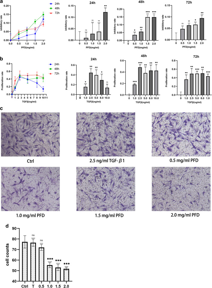
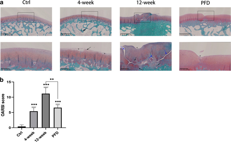
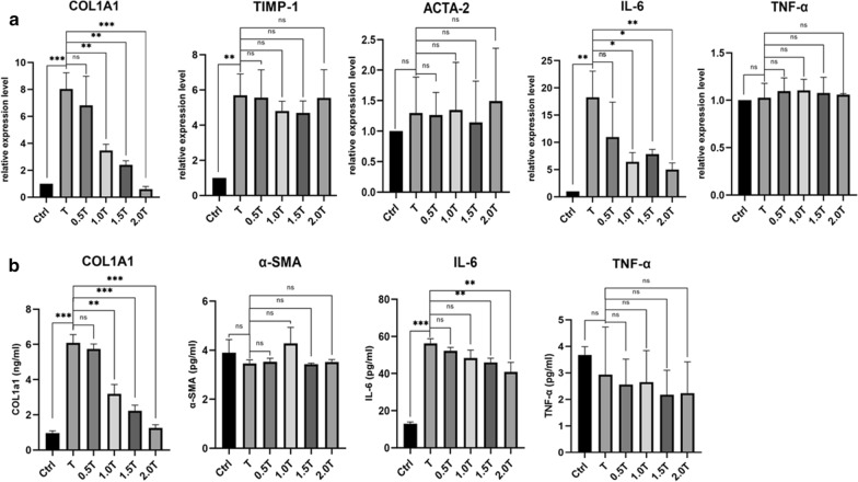
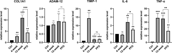
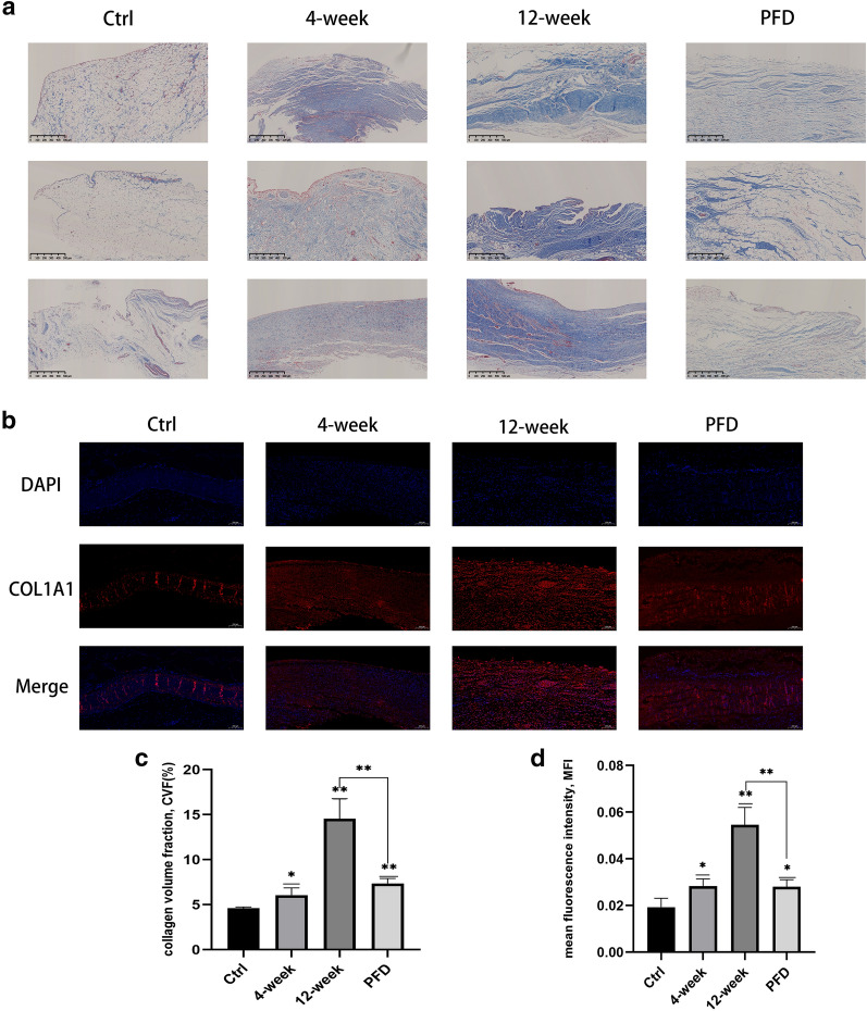
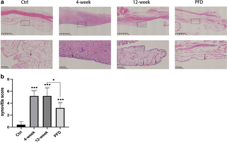
Proliferation of human fibroblast-like synoviocytes (FLSs) after treatment with TGF-β1 or PFD was evaluated using a Cell Counting Kit-8 assay and their migration using a Transwell assay. The expression of fibrosis-related genes (COL1A1, TIMP-1, and ACTA-2) and those related to inflammation (IL-6 and TNF-α) was quantified by real-time quantitative PCR. The protein expression levels of COL1A1, α-SMA (coded by ACTA-2), IL-6 and TNF-α were measured by enzyme-linked immunosorbent assay. A rabbit model of OA was established and then PFD was administered by gavage. The expression of genes related to fibrosis (COL1A1, TIMP-1, and ADAM-12) and inflammation (IL-6 and TNF-α) was measured using RNA extracted from the synovium. Synovial tissue was examined histologically after staining with H&E, Masson's trichrome, and immunofluorescence. Synovitis scores, the volume fraction of collagen, and mean fluorescence intensity were calculated. Degeneration of articular cartilage was analyzed using a Safranin O-fast green stain and OARSI grading.
The proliferation of FLSs was greatest when induced with 2.5 ng/ml TGF-β1 although it did not promote their migration. Therefore, 2.5 ng/ml TGF-β1 was used to stimulate the FLSs and evaluate the effects of PFD, which inhibited the migration of FLSs at concentrations as low as 1.0 mg/ml. PFD decreased the expression of COL1A1 while TGF-β1 increased both mRNA and protein expression levels of IL-6 but had no effect on α-SMA or TNF-α expression. PFD decreased mRNA expression levels of COL1A1, IL-6, and TNF-α in vivo. H&E staining and synovitis scores indicated that PFD reduced synovial inflammation, while Masson's trichrome and immunofluorescence staining suggested that PFD decreased synovial fibrosis. Safranin O-Fast Green staining and the OARSI scores demonstrated that PFD delayed the progression of OA.
PFD attenuated synovial fibrosis and inflammation, and postponed the progression of osteoarthritis in a modified Hulth model of OA in rabbits, which was related to its anti-fibrotic and anti-inflammatory properties.
骨关节炎(OA)是一种累及整个关节的疾病,包括滑膜纤维化和炎症。滑膜的病理变化可加速OA的进展。吡非尼酮(PFD)是一种具有额外抗炎特性的强效抗纤维化药物。然而,PFD对OA的影响尚不清楚。
使用细胞计数试剂盒-8法评估用转化生长因子-β1(TGF-β1)或PFD处理后人成纤维样滑膜细胞(FLS)的增殖情况,并用Transwell法评估其迁移情况。通过实时定量PCR定量纤维化相关基因(COL1A1、TIMP-1和ACTA-2)以及炎症相关基因(IL-6和TNF-α)的表达。通过酶联免疫吸附测定法测量COL1A1、α-平滑肌肌动蛋白(由ACTA-2编码)、IL-6和TNF-α的蛋白表达水平。建立OA兔模型,然后通过灌胃给予PFD。使用从滑膜中提取的RNA测量纤维化相关基因(COL1A1、TIMP-1和ADAM-12)以及炎症相关基因(IL-6和TNF-α)的表达。用苏木精-伊红(H&E)、Masson三色染色和免疫荧光染色后对滑膜组织进行组织学检查。计算滑膜炎评分、胶原蛋白体积分数和平均荧光强度。使用番红O-固绿染色和骨关节炎研究学会国际(OARSI)分级分析关节软骨退变情况。
用2.5 ng/ml TGF-β1诱导时FLS的增殖最为显著,尽管它不促进其迁移。因此,使用2.5 ng/ml TGF-β1刺激FLS并评估PFD的作用,PFD在低至1.0 mg/ml的浓度下即可抑制FLS的迁移。PFD降低COL1A1的表达,而TGF-β1增加IL-6的mRNA和蛋白表达水平,但对α-平滑肌肌动蛋白或TNF-α表达无影响。PFD在体内降低COL1A1、IL-6和TNF-α的mRNA表达水平。H&E染色和滑膜炎评分表明PFD减轻了滑膜炎症,而Masson三色染色和免疫荧光染色表明PFD减少了滑膜纤维化。番红O-固绿染色和OARSI评分表明PFD延缓了OA的进展。
在改良的兔OA Hulth模型中,PFD减轻了滑膜纤维化和炎症,并延缓了骨关节炎的进展,这与其抗纤维化和抗炎特性有关。