Liao Bo, Guan Mengtong, Tan Qiaoyan, Wang Gailan, Zhang Ruobin, Huang Junlan, Liu Mi, Chen Hong, Li Kaiting, Bai Dingqun, Zhu Ying
Department of Rehabilitation Medicine, The First Affiliated Hospital of Chongqing Medical University, Chongqing, 400010, China.
Department of Rehabilitation Medicine, Chengdu Women's and Children's Central Hospital, Chengdu, Sichuan province, 610091, China.
J Orthop Translat. 2021 Sep 20;30:41-50. doi: 10.1016/j.jot.2021.08.002. eCollection 2021 Sep.
Synovial fibrosis is a characteristic symptom of osteoarthritis (OA), which is closely associated with joint pain and stiffness. Previous studies have reported that low-intensity pulsed ultrasound (LIPUS) can alleviate cartilage degradation in OA. However, the functions and mechanisms of LIPUS in OA synovial fibrosis are still unknown.
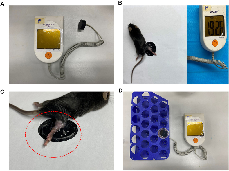
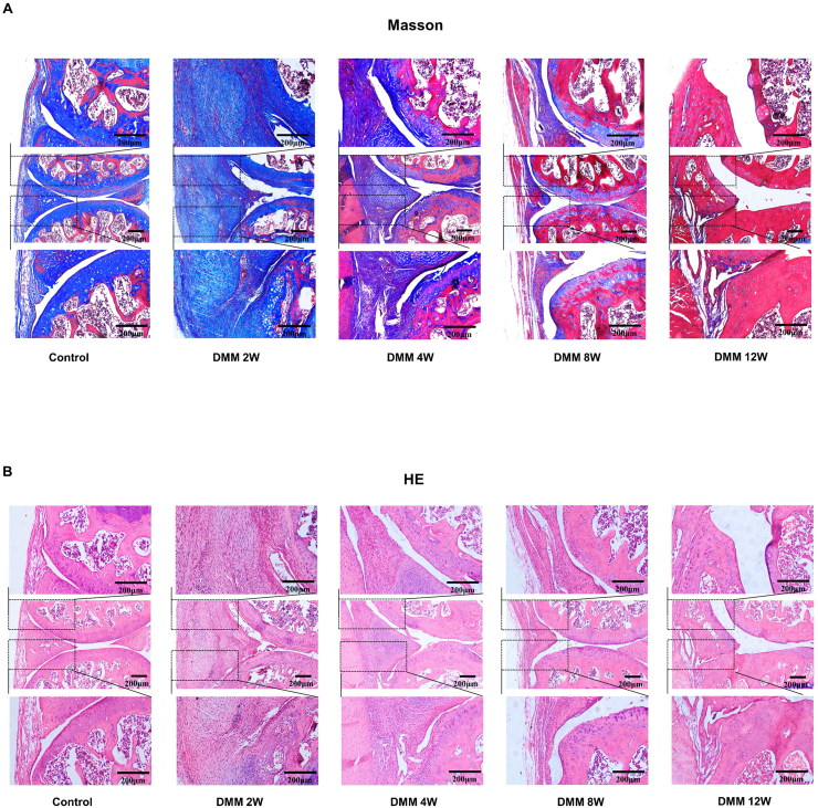
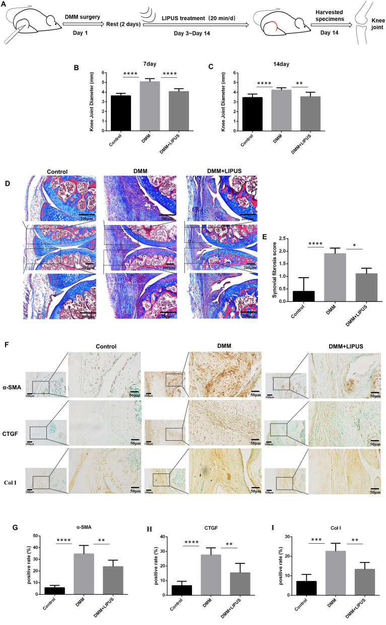
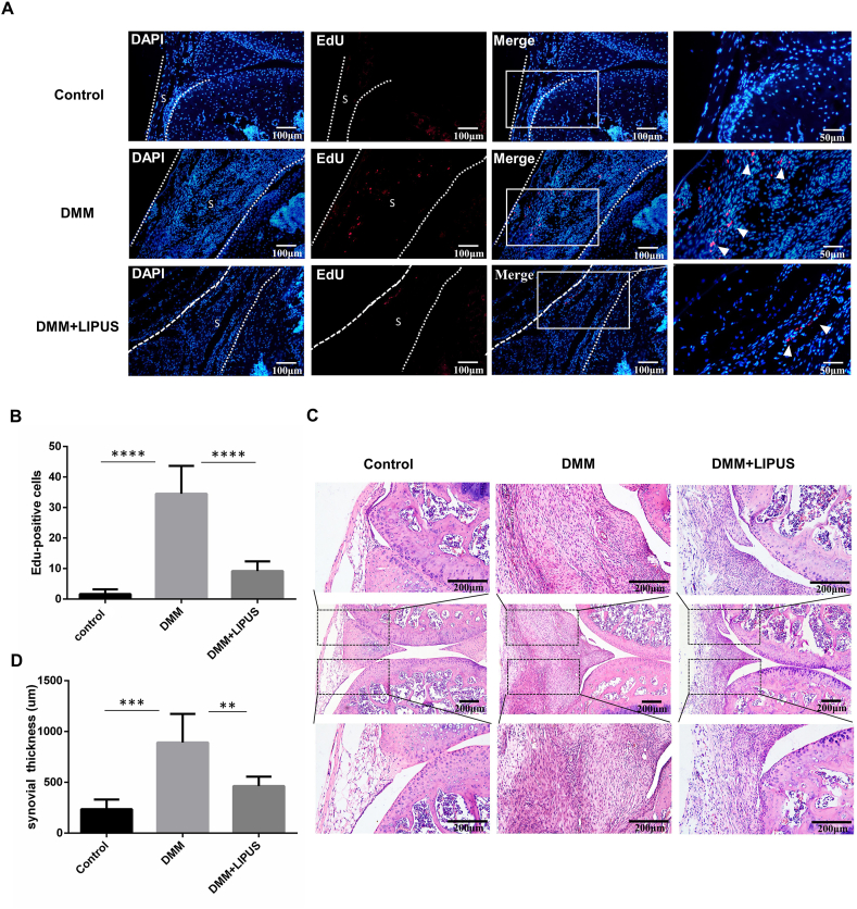
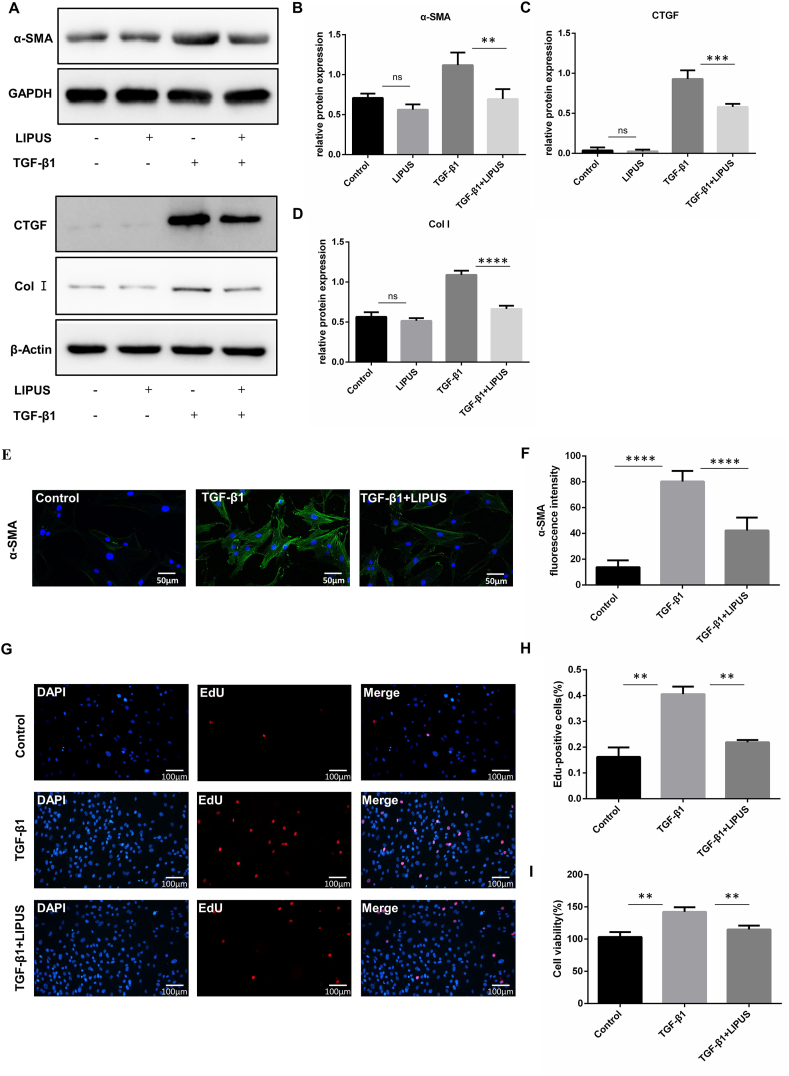
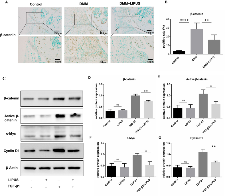
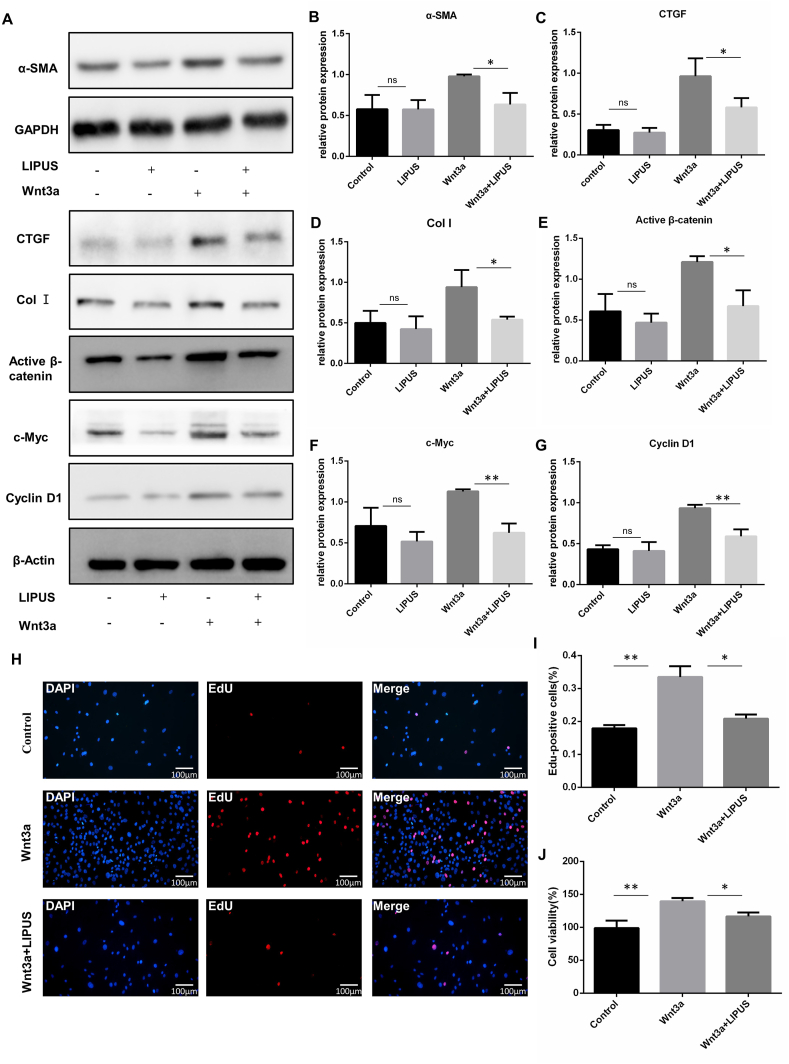
The destabilization of the medial meniscus (DMM) mouse model of OA was established in C57 male mice and fibroblast-like synoviocytes (FLS) were isolated from synovial tissue of OA patients. The knee joint diameter, Masson's trichrome (MT) and Hematoxylin-eosin (HE) staining were used to evaluate synovial fibrosis and hyperplasia. The Immunohistochemistry (IHC) staining was performed to detected the expression of synovial fibrosis makers and the activation of Wnt/β-catenin signaling . FLS were treated with TGF-β1 to serve as an model of synovial fibrosis, Wnt3a was used to activate the Wnt/β-catenin signaling in cells. Cell proliferation was detected by using EdU assay, cell viability was performed by CCK8 assay. The protein levels of α-SMA, CTGF, Col Ⅰ, β-catenin, active β-catenin, c-Myc and cyclin D1 were examined by western blot and immunofluorescence staining.
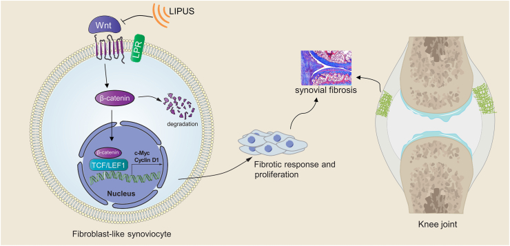
Two weeks after the LIPUS treatment, the synovial fibrosis, synovial hyperplasia and synoviocyte proliferation in the DMM model were significantly decreased. , LIPUS directly inhibited the TGF-β1-induced fibrotic response and proliferation of FLS. Meanwhile, LIPUS suppressed Wnt/β-catenin signaling in the synovium of DMM mice and cultured FLS. More importantly, we found that the synovial fibrosis makers, Wnt/β-catenin pathway downstream proteins and FLS proliferation were significantly decreased in Wnt3a-stimulated FLS following LIPUS treatment.
Our results present a novel role of LIPUS in OA-related synovial fibrosis, which is associated with its ability to repress Wnt/β-catenin signaling in FLS.
This study provides new insight into the clinical application of LIPUS as a therapeutic option to manage synovial fibrosis in OA.
滑膜纤维化是骨关节炎(OA)的一个特征性症状,与关节疼痛和僵硬密切相关。既往研究报道,低强度脉冲超声(LIPUS)可减轻OA中的软骨降解。然而,LIPUS在OA滑膜纤维化中的作用及机制仍不清楚。
在C57雄性小鼠中建立OA的内侧半月板不稳定(DMM)小鼠模型,并从OA患者的滑膜组织中分离出成纤维样滑膜细胞(FLS)。采用膝关节直径、Masson三色染色(MT)和苏木精-伊红染色(HE)评估滑膜纤维化和增生情况。进行免疫组织化学(IHC)染色以检测滑膜纤维化标志物的表达及Wnt/β-连环蛋白信号通路的激活情况。用转化生长因子-β1(TGF-β1)处理FLS以建立滑膜纤维化模型,用Wnt3a激活细胞中的Wnt/β-连环蛋白信号通路。采用EdU检测法检测细胞增殖,用CCK8检测法检测细胞活力。通过蛋白质印迹法和免疫荧光染色检测α-平滑肌肌动蛋白(α-SMA)、结缔组织生长因子(CTGF)、Ⅰ型胶原(Col Ⅰ)、β-连环蛋白、活性β-连环蛋白、c-Myc和细胞周期蛋白D1的蛋白水平。
LIPUS治疗两周后,DMM模型中的滑膜纤维化、滑膜增生和滑膜细胞增殖明显减少。此外,LIPUS直接抑制TGF-β1诱导的FLS纤维化反应和增殖。同时LIPUS抑制DMM小鼠滑膜和培养的FLS中的Wnt/β-连环蛋白信号通路。更重要的是,我们发现LIPUS处理后的Wnt3a刺激的FLS中,滑膜纤维化标志物、Wnt/β-连环蛋白通路下游蛋白和FLS增殖明显减少。
我们的结果揭示了LIPUS在OA相关滑膜纤维化中的新作用,这与其抑制FLS中Wnt/β-连环蛋白信号通路的能力有关。
本研究为LIPUS作为治疗OA滑膜纤维化的临床应用提供了新的见解。