Department of Bioengineering, University of Illinois at Urbana-Champaign, Urbana, IL 61801, USA.
Nick Holonyak Jr. Micro and Nanotechnology Laboratory, University of Illinois at Urbana-Champaign, Urbana, IL 61801, USA.
Sci Adv. 2021 Apr 23;7(17). doi: 10.1126/sciadv.abc1323. Print 2021 Apr.
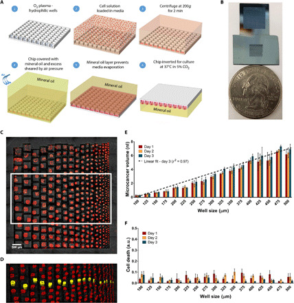
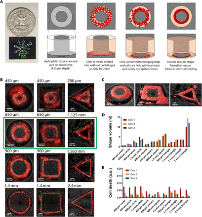
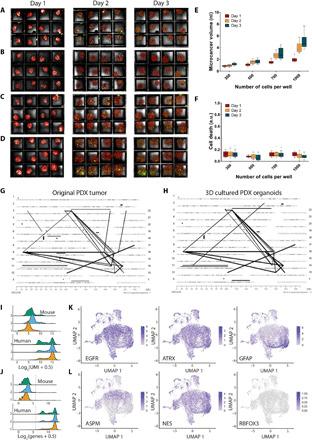
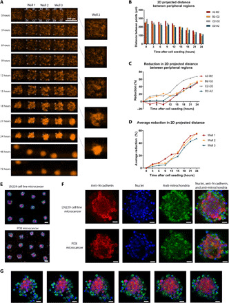
Existing three-dimensional (3D) culture techniques are limited by trade-offs between throughput, capacity for high-resolution imaging in living state, and geometric control. Here, we introduce a modular microscale hanging drop culture where simple design elements allow high replicates for drug screening, direct on-chip real-time or high-resolution confocal microscopy, and geometric control in 3D. Thousands of spheroids can be formed on our microchip in a single step and without any selective pressure from specific matrices. Microchip cultures from human LN229 glioblastoma and patient-derived mouse xenograft cells retained genomic alterations of originating tumors based on mate pair sequencing. We measured response to drugs over time with real-time microscopy on-chip. Last, by engineering droplets to form predetermined geometric shapes, we were able to manipulate the geometry of cultured cell masses. These outcomes can enable broad applications in advancing personalized medicine for cancer and drug discovery, tissue engineering, and stem cell research.
现有的三维(3D)培养技术在通量、活细胞高分辨率成像能力和几何形状可控性之间存在权衡。本文介绍了一种模块化微尺度悬滴培养方法,其简单的设计元素可实现高通量药物筛选、直接在芯片上进行实时或高分辨率共聚焦显微镜检测,以及 3D 几何形状可控性。在单个步骤中,我们可以在微芯片上形成数千个球体,而无需特定基质的任何选择性压力。基于配对末端测序,来自人 LN229 神经胶质瘤和患者来源的小鼠异种移植细胞的微芯片培养物保留了原始肿瘤的基因组改变。我们通过实时显微镜在芯片上测量了随时间的药物反应。最后,通过设计液滴形成预定的几何形状,我们能够操纵培养细胞块的几何形状。这些结果可以广泛应用于推进癌症和药物发现、组织工程和干细胞研究的个性化医疗。