Department of Bioengineering, University of Illinois at Urbana-Champaign, Champaign, IL, 61801, USA.
Micro and Nanotechnology Laboratory, University of Illinois at Urbana-Champaign, Champaign, IL, 61801, USA.
Nat Commun. 2018 Jan 15;9(1):202. doi: 10.1038/s41467-017-02623-9.
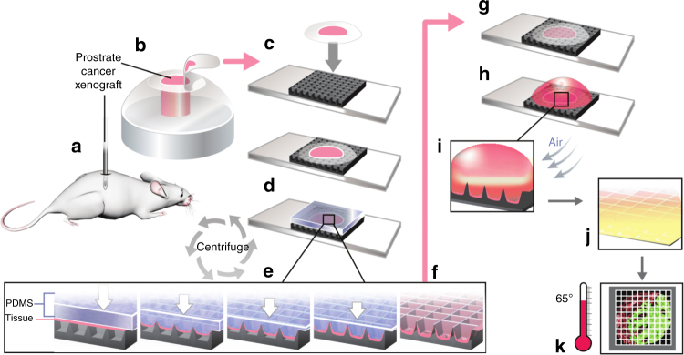
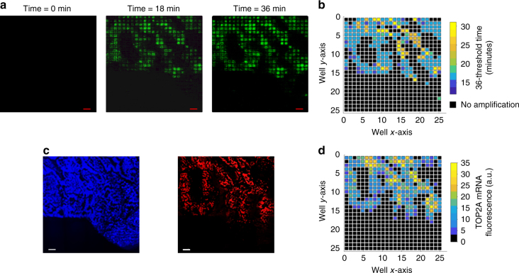
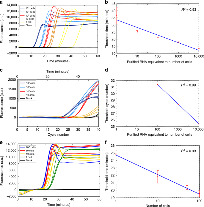
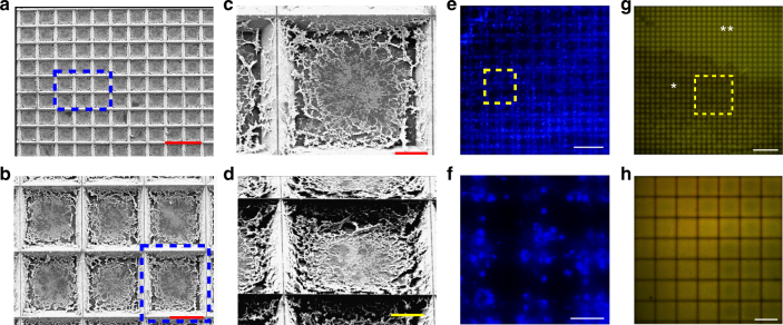
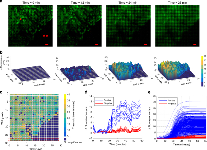
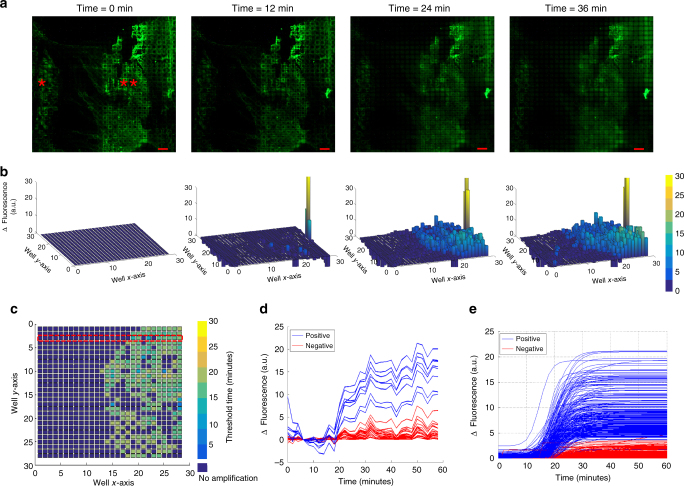
Here, we present a technique that performs on-chip picoliter real-time reverse transcriptase loop mediated isothermal amplification (RT-LAMP) reactions on a histological tissue section without any analyte purification while preserving the native spatial location of the nucleic acid molecules. We demonstrate this method by amplifying TOP2A messenger RNA (mRNA) in a prostate cancer xenograft with 100 µm spatial resolution and by visualizing the variation in threshold time of amplification across the tissue. The on-chip reaction was validated by mRNA fluorescence in situ hybridization (mFISH) from cells in the tissue section. The entire process, from tissue loading on microchip to results from RT-LAMP can be carried out in less than 2 h. We anticipate that this technique, with its ease of use, fast turnaround, and quantitative molecular outputs, would become an invaluable tissue analysis tool for researchers and clinicians in the biomedical arena.
在这里,我们提出了一种技术,可在不进行任何分析物纯化的情况下,在组织切片上进行片上皮升实时逆转录环介导等温扩增(RT-LAMP)反应,同时保留核酸分子的天然空间位置。我们通过以 100μm 的空间分辨率扩增前列腺癌异种移植物中的 TOP2A 信使 RNA(mRNA),并通过可视化组织中扩增的阈值时间变化来证明这种方法。通过组织切片中的细胞 mRNA 荧光原位杂交(mFISH)验证了芯片上的反应。从组织加载到 RT-LAMP 的结果,整个过程可以在不到 2 小时内完成。我们预计,这种技术具有使用简便、周转快速和定量分子输出等特点,将成为生物医学领域研究人员和临床医生非常有价值的组织分析工具。