Department of Radiology, University of Cambridge School of Clinical Medicine, University of Cambridge, Cambridge Biomedical Campus, Box 218, Cambridge, CB2 0QQ, UK.
MMIV, Department of Radiology, Haukeland University Hospital, Bergen, Norway.
Sci Rep. 2021 Apr 23;11(1):8857. doi: 10.1038/s41598-021-87857-w.
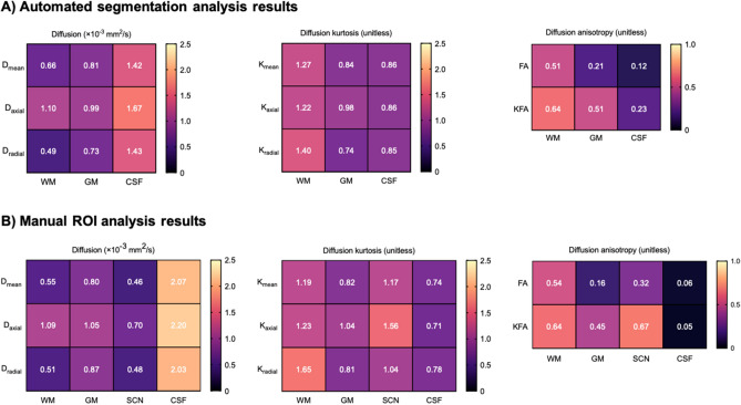
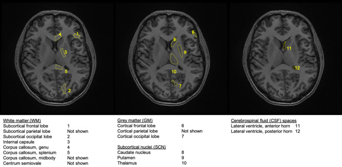
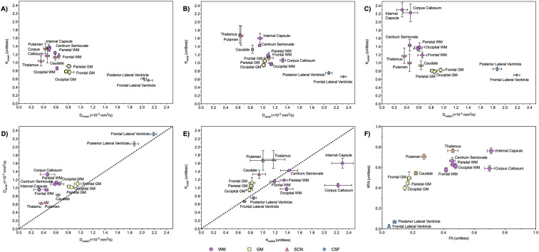
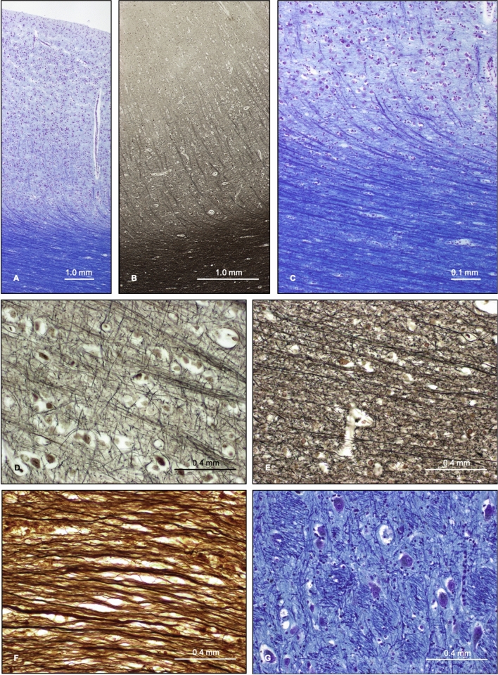
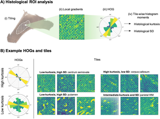
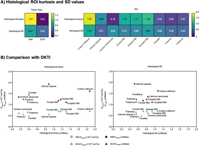
Measurements of water diffusion with MRI have been used as a biomarker of tissue microstructure and heterogeneity. In this study, diffusion kurtosis tensor imaging (DKTI) of the brain was undertaken in 10 healthy volunteers at a clinical field strength of 3 T. Diffusion and kurtosis metrics were measured in regions-of-interest on the resulting maps and compared with quantitative analysis of normal post-mortem tissue histology from separate age-matched donors. White matter regions showed low diffusion (0.60 ± 0.04 × 10 mm/s) and high kurtosis (1.17 ± 0.06), consistent with a structured heterogeneous environment comprising parallel neuronal fibres. Grey matter showed intermediate diffusion (0.80 ± 0.02 × 10 mm/s) and kurtosis (0.82 ± 0.05) values. An important finding is that the subcortical regions investigated (thalamus, caudate and putamen) showed similar diffusion and kurtosis properties to white matter. Histological staining of the subcortical nuclei demonstrated that the predominant grey matter was permeated by small white matter bundles, which could account for the similar kurtosis to white matter. Quantitative histological analysis demonstrated higher mean tissue kurtosis and vector standard deviation values for white matter (1.08 and 0.81) compared to the subcortical regions (0.34 and 0.59). Mean diffusion on DKTI was positively correlated with tissue kurtosis (r = 0.82, p < 0.05) and negatively correlated with vector standard deviation (r = -0.69, p < 0.05). This study demonstrates how DKTI can be used to study regional structural variations in the cerebral tissue microenvironment and could be used to probe microstructural changes within diseased tissue in the future.
磁共振成像(MRI)的水分子扩散测量已被用作组织微观结构和异质性的生物标志物。在这项研究中,在临床 3T 场强下对 10 名健康志愿者的大脑进行了扩散峰度张量成像(DKTI)。在所得图谱的感兴趣区域中测量了扩散和峰度指标,并与来自单独年龄匹配供体的定量分析正常尸检组织组织学进行了比较。白质区域显示出低扩散(0.60 ± 0.04×10mm/s)和高峰度(1.17 ± 0.06),与包含平行神经元纤维的结构化异质环境一致。灰质显示出中等扩散(0.80 ± 0.02×10mm/s)和峰度(0.82 ± 0.05)值。一个重要的发现是,研究的皮质下区域(丘脑、尾状核和壳核)显示出与白质相似的扩散和峰度特性。皮质下核的组织学染色表明,主要的灰质被小的白质束渗透,这可以解释与白质相似的峰度。定量组织学分析表明,白质的平均组织峰度和向量标准差值较高(1.08 和 0.81),而皮质下区域的峰度和向量标准差值较低(0.34 和 0.59)。DKTI 上的平均扩散与组织峰度呈正相关(r=0.82,p<0.05),与向量标准差呈负相关(r=-0.69,p<0.05)。这项研究表明,DKTI 如何用于研究大脑组织微环境中的区域结构变化,并可用于未来探测病变组织内的微观结构变化。