Center for Neuroscience Imaging Research (CNIR), Institute for Basic Science (IBS), Suwon 16419, Republic of Korea.
Department of Biomedical Engineering, Sungkyunkwan University, Suwon 16419, Republic of Korea.
Cereb Cortex. 2021 Jul 29;31(9):4053-4067. doi: 10.1093/cercor/bhab068.
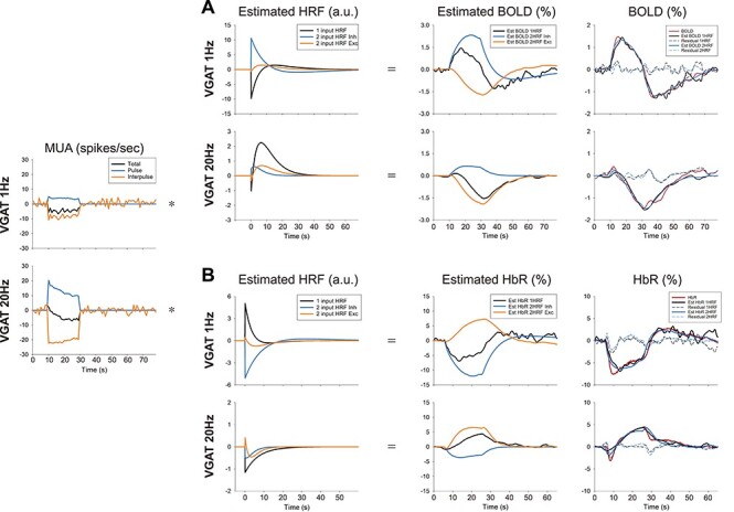
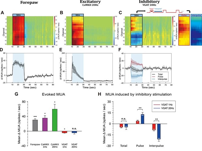
The BOLD fMRI response in the cortex is often assumed to reflect changes in excitatory neural activity. However, the contribution of inhibitory neurons to BOLD fMRI is unclear. Here, the role of inhibitory and excitatory activity was examined using multimodal approaches: electrophysiological recording, 15.2 T fMRI, optical intrinsic signal imaging, and modeling. Inhibitory and excitatory neuronal activity in the somatosensory cortex were selectively modulated by 20-s optogenetic stimulation of VGAT-ChR2 and CaMKII-ChR2 mice, respectively. Somatosensory stimulation and optogenetic stimulation of excitatory neurons induced positive BOLD responses in the somatosensory network, whereas stimulation of inhibitory neurons produced biphasic responses at the stimulation site, initial positive and later negative BOLD signals, and negative BOLD responses at downstream sites. When the stimulation duration was reduced to 5 s, the hemodynamic response of VGAT-ChR2 mice to optogenetic stimulation was only positive. Lastly, modeling performed from neuronal and hemodynamic data shows that the hemodynamic response function (HRF) of excitatory neurons is similar across different conditions, whereas the HRF of inhibitory neurons is highly sensitive to stimulation frequency and peaks earlier than that of excitatory neurons. Our study provides insights into the neurovascular coupling of excitatory and inhibitory neurons and the interpretation of BOLD fMRI signals.
皮层中的 BOLD fMRI 反应通常被认为反映了兴奋性神经活动的变化。然而,抑制性神经元对 BOLD fMRI 的贡献尚不清楚。在这里,使用多模态方法研究了抑制性和兴奋性活动的作用:电生理记录、15.2T fMRI、光学内源信号成像和建模。通过分别对 VGAT-ChR2 和 CaMKII-ChR2 小鼠进行 20 秒光遗传学刺激,选择性调节感觉皮层中的抑制性和兴奋性神经元活动。感觉刺激和兴奋性神经元的光遗传学刺激在感觉网络中诱导了正的 BOLD 反应,而抑制性神经元的刺激在刺激部位产生双相反应,最初为正 BOLD 信号,随后为负 BOLD 信号,下游部位为负 BOLD 反应。当刺激持续时间缩短至 5 秒时,VGAT-ChR2 小鼠对光遗传学刺激的血液动力学反应仅为正。最后,从神经元和血液动力学数据进行的建模表明,兴奋性神经元的血液动力学反应功能(HRF)在不同条件下相似,而抑制性神经元的 HRF 对刺激频率高度敏感,并且比兴奋性神经元更早达到峰值。我们的研究提供了对兴奋性和抑制性神经元的神经血管偶联以及 BOLD fMRI 信号解释的深入了解。