• 文献检索
  • 文档翻译
  • 深度研究
  • 学术资讯
  • Suppr Zotero 插件Zotero 插件
  • 邀请有礼
  • 套餐&价格
  • 历史记录
应用&插件
Suppr Zotero 插件Zotero 插件浏览器插件Mac 客户端Windows 客户端微信小程序
定价
高级版会员购买积分包购买API积分包
服务
文献检索文档翻译深度研究API 文档MCP 服务
关于我们
关于 Suppr公司介绍联系我们用户协议隐私条款
关注我们

Suppr 超能文献

核心技术专利:CN118964589B侵权必究
粤ICP备2023148730 号-1Suppr @ 2026

文献检索

告别复杂PubMed语法,用中文像聊天一样搜索,搜遍4000万医学文献。AI智能推荐,让科研检索更轻松。

立即免费搜索

文件翻译

保留排版,准确专业,支持PDF/Word/PPT等文件格式,支持 12+语言互译。

免费翻译文档

深度研究

AI帮你快速写综述,25分钟生成高质量综述,智能提取关键信息,辅助科研写作。

立即免费体验

Ki 或非 Ki:重新评估 Ki-67 在 T 细胞分析中的应用和潜力。

To Ki or Not to Ki: Re-Evaluating the Use and Potentials of Ki-67 for T Cell Analysis.

机构信息

Institute of Molecular Biology and Pathology, National Research Council of Italy (CNR), Rome, Italy.

Department of Medical and Surgical Sciences for Children and Adults, University of Modena and Reggio Emilia, Modena, Italy.

出版信息

Front Immunol. 2021 Apr 9;12:653974. doi: 10.3389/fimmu.2021.653974. eCollection 2021.

DOI:10.3389/fimmu.2021.653974
PMID:33897702
原文链接:https://pmc.ncbi.nlm.nih.gov/articles/PMC8062736/
Abstract

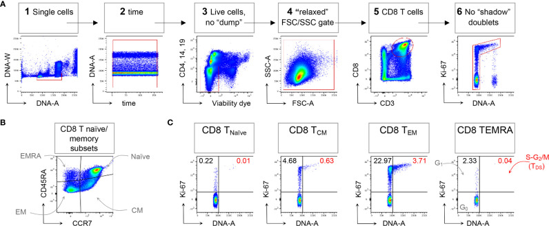
This study discusses substantive advances in T cell proliferation analysis, with the aim to provoke a re-evaluation of the generally-held view that Ki-67 is a reliable proliferation marker , and to offer a more sensitive and effective method for T cell cycle analysis, with informative examples in mouse and human settings. We summarize recent experimental work from our labs showing that, by Ki-67/DNA dual staining and refined flow cytometric methods, we were able to identify T cells in the S-G/M phases of the cell-cycle in the peripheral blood (collectively termed "T Double S" for T cells in S-phase : in short "T" cells). Without our refinement, such cells may be excluded from conventional lymphocyte analyses. Specifically, we analyzed clonal expansion of antigen-specific CD8 T cells in vaccinated mice, and demonstrated the potential of T cells to reflect immune dynamics in human blood samples from healthy donors, and patients with type 1 diabetes, infectious mononucleosis, and COVID-19. The Ki-67/DNA dual staining, or T assay, provides a reliable approach by which human peripheral blood can be used to reflect the dynamics of human lymphocytes, rather than providing mere steady-state phenotypic snapshots. The method does not require highly sophisticated "-omics" capabilities, so it should be widely-applicable to health care in diverse settings. Furthermore, our results argue that the T assay can provide a window on immune dynamics in extra-lymphoid tissues, a long-sought potential of peripheral blood monitoring, for example in relation to organ-specific autoimmune diseases and infections, and cancer immunotherapy.

摘要

本研究探讨了 T 细胞增殖分析的实质性进展,旨在重新评估普遍认为 Ki-67 是可靠增殖标志物的观点,并提供一种更敏感和有效的 T 细胞周期分析方法,同时提供了在小鼠和人类研究中的实例。我们总结了最近来自我们实验室的实验工作,表明通过 Ki-67/DNA 双重染色和改良的流式细胞术方法,我们能够在外周血中识别处于细胞周期 S-G/M 期的 T 细胞(统称为 S 期的 T 双 S:简称“T”细胞)。如果没有我们的改进,这些细胞可能会从常规淋巴细胞分析中排除。具体来说,我们分析了疫苗接种小鼠中抗原特异性 CD8 T 细胞的克隆扩增,并证明了 T 细胞在健康供体和 1 型糖尿病、传染性单核细胞增多症和 COVID-19 患者的人类血液样本中反映免疫动态的潜力。Ki-67/DNA 双重染色或 T 检测提供了一种可靠的方法,可以使用人类外周血来反映人类淋巴细胞的动态,而不仅仅是提供静止状态的表型快照。该方法不需要高度复杂的“组学”能力,因此应该在不同环境下广泛应用于医疗保健。此外,我们的结果表明,T 检测可以提供一个观察淋巴外组织免疫动态的窗口,这是外周血监测长期以来的一个潜在需求,例如与器官特异性自身免疫疾病和感染以及癌症免疫治疗有关。

https://cdn.ncbi.nlm.nih.gov/pmc/blobs/5a34/8062736/8f1a81fc3dbc/fimmu-12-653974-g001.jpg

相似文献

1
To Ki or Not to Ki: Re-Evaluating the Use and Potentials of Ki-67 for T Cell Analysis.Ki 或非 Ki:重新评估 Ki-67 在 T 细胞分析中的应用和潜力。
Front Immunol. 2021 Apr 9;12:653974. doi: 10.3389/fimmu.2021.653974. eCollection 2021.
2
Tracking immunodynamics by identification of S-G/M-phase T cells in human peripheral blood.通过鉴定人外周血中的 S-G/M 期 T 细胞来跟踪免疫动力学。
J Autoimmun. 2020 Aug;112:102466. doi: 10.1016/j.jaut.2020.102466. Epub 2020 May 12.
3
A DNA/Ki67-Based Flow Cytometry Assay for Cell Cycle Analysis of Antigen-Specific CD8 T Cells in Vaccinated Mice.基于 DNA/Ki67 的流式细胞术检测疫苗接种小鼠中抗原特异性 CD8 T 细胞的细胞周期分析。
J Vis Exp. 2021 Jan 5(167). doi: 10.3791/61867.
4
Repurposing CD8 T cell immunity against SARS-CoV-2 for cancer immunotherapy: a positive aspect of the COVID-19 pandemic?重新利用针对 SARS-CoV-2 的 CD8 T 细胞免疫应答进行癌症免疫治疗:COVID-19 大流行的一个积极方面?
Oncoimmunology. 2020 Jul 16;9(1):1794424. doi: 10.1080/2162402X.2020.1794424.
5
T Cell Phenotyping in Individuals Hospitalized with COVID-19.COVID-19 住院患者的 T 细胞表型分析。
J Immunol. 2021 Apr 1;206(7):1478-1482. doi: 10.4049/jimmunol.2001034. Epub 2021 Feb 8.
6
Antigen-specific CD8 T cells in cell cycle circulate in the blood after vaccination.接种疫苗后,处于细胞周期中的抗原特异性 CD8 T 细胞会在血液中循环。
Scand J Immunol. 2019 Feb;89(2):e12735. doi: 10.1111/sji.12735. Epub 2019 Jan 15.
7
IL-7 in SARS-CoV-2 Infection and as a Potential Vaccine Adjuvant.白细胞介素 7 在 SARS-CoV-2 感染中的作用及其作为潜在疫苗佐剂的潜力。
Front Immunol. 2021 Sep 17;12:737406. doi: 10.3389/fimmu.2021.737406. eCollection 2021.
8
A single dose of self-transcribing and replicating RNA-based SARS-CoV-2 vaccine produces protective adaptive immunity in mice.一剂自我转录和复制的基于 RNA 的 SARS-CoV-2 疫苗可在小鼠中产生保护性适应性免疫。
Mol Ther. 2021 Jun 2;29(6):1970-1983. doi: 10.1016/j.ymthe.2021.04.001. Epub 2021 Apr 5.
9
[Vaccination immunology in SARS-CoV-2].[新型冠状病毒肺炎中的疫苗接种免疫学]
Ugeskr Laeger. 2021 Mar 15;183(11).
10
SARS-CoV-2-specific T cell memory is sustained in COVID-19 convalescent patients for 10 months with successful development of stem cell-like memory T cells.SARS-CoV-2 特异性 T 细胞记忆在 COVID-19 恢复期患者中持续存在 10 个月,成功发育出干细胞样记忆 T 细胞。
Nat Commun. 2021 Jun 30;12(1):4043. doi: 10.1038/s41467-021-24377-1.

引用本文的文献

1
Gut IgA-antibody secreting cells segregate into four Blimp1+ subsets based on differential expression of IgA and Ki-67 and are retained following prolonged αCD20 B cell depletion in mice.肠道分泌IgA抗体的细胞根据IgA和Ki-67的差异表达分为四个Blimp1+亚群,并且在小鼠长期αCD20 B细胞耗竭后仍保留。
J Immunol. 2025 Apr 1;214(4):780-794. doi: 10.1093/jimmun/vkae046.
2
Proliferating CLL cells express high levels of CXCR4 and CD5.增殖性慢性淋巴细胞白血病细胞表达高水平的CXCR4和CD5。
Hemasphere. 2024 Dec 17;8(12):e70064. doi: 10.1002/hem3.70064. eCollection 2024 Dec.
3
Macrophage subtypes inhibit breast cancer proliferation in culture.

本文引用的文献

1
Acute Immune Signatures and Their Legacies in Severe Acute Respiratory Syndrome Coronavirus-2 Infected Cancer Patients.严重急性呼吸综合征冠状病毒 2 感染癌症患者的急性免疫特征及其后果。
Cancer Cell. 2021 Feb 8;39(2):257-275.e6. doi: 10.1016/j.ccell.2021.01.001. Epub 2021 Jan 5.
2
Fate Mapping Quantifies the Dynamics of B Cell Development and Activation throughout Life.命运图谱定量描绘了 B 细胞在整个生命过程中的发育和激活动态。
Cell Rep. 2020 Nov 17;33(7):108376. doi: 10.1016/j.celrep.2020.108376.
3
Chromosome clustering by Ki-67 excludes cytoplasm during nuclear assembly.
巨噬细胞亚型在培养中抑制乳腺癌增殖。
Mol Biol Cell. 2025 Jan 1;36(1):br2. doi: 10.1091/mbc.E24-06-0241. Epub 2024 Nov 27.
4
Macrophage subtypes inhibit breast cancer proliferation in culture.巨噬细胞亚型在培养中抑制乳腺癌增殖。
bioRxiv. 2024 Jun 1:2024.06.01.596963. doi: 10.1101/2024.06.01.596963.
5
Single-Cell RNA Sequencing Reveals Immuno-Oncology Characteristics of Tumor-Infiltrating T Lymphocytes in Photodynamic Therapy-Treated Colorectal Cancer Mouse Model.单细胞RNA测序揭示光动力疗法治疗的结直肠癌小鼠模型中肿瘤浸润性T淋巴细胞的免疫肿瘤学特征
Int J Mol Sci. 2023 Sep 10;24(18):13913. doi: 10.3390/ijms241813913.
6
New Flow Cytometric Methods for Monitoring STAT5 Signaling Reveal Responses to SARS-CoV-2 Antigen-Specific Stimulation in FOXP3+ Regulatory T Cells also in Patients with Advanced Chronic Lymphocytic Leukemia.新型流式细胞术方法监测 STAT5 信号通路揭示了 SARS-CoV-2 抗原特异性刺激在 FOXP3+调节性 T 细胞中的反应,也揭示了晚期慢性淋巴细胞白血病患者中的反应。
Biosensors (Basel). 2023 May 11;13(5):539. doi: 10.3390/bios13050539.
7
The Epigenetic Reader Protein SP140 Regulates Dendritic Cell Activation, Maturation and Tolerogenic Potential.表观遗传阅读蛋白SP140调节树突状细胞的激活、成熟和致耐受性潜能。
Curr Issues Mol Biol. 2023 May 11;45(5):4228-4245. doi: 10.3390/cimb45050269.
8
The SEB1741 Aptamer Is an Efficient Tool for Blocking CD4+ T Cell Activation Induced by Staphylococcal Enterotoxin B.SEB1741 适体是一种有效的工具,可阻断葡萄球菌肠毒素 B 诱导的 CD4+T 细胞活化。
Molecules. 2023 Apr 14;28(8):3480. doi: 10.3390/molecules28083480.
9
Downregulation of MALAT1 is a hallmark of tissue and peripheral proliferative T cells in COVID-19.MALAT1 的下调是 COVID-19 中组织和外周增殖性 T 细胞的一个标志。
Clin Exp Immunol. 2023 Jun 5;212(3):262-275. doi: 10.1093/cei/uxad034.
10
Low-dose IL-2 reduces IL-21 T cell frequency and induces anti-inflammatory gene expression in type 1 diabetes.低剂量白介素 2 减少 1 型糖尿病中白介素 21 T 细胞频率并诱导抗炎基因表达。
Nat Commun. 2022 Nov 28;13(1):7324. doi: 10.1038/s41467-022-34162-3.
Ki-67 将染色体聚类排除在核组装过程中的细胞质之外。
Nature. 2020 Nov;587(7833):285-290. doi: 10.1038/s41586-020-2672-3. Epub 2020 Sep 2.
4
A dynamic COVID-19 immune signature includes associations with poor prognosis.一个动态的 COVID-19 免疫特征包括与预后不良的关联。
Nat Med. 2020 Oct;26(10):1623-1635. doi: 10.1038/s41591-020-1038-6. Epub 2020 Aug 17.
5
Tracking immunodynamics by identification of S-G/M-phase T cells in human peripheral blood.通过鉴定人外周血中的 S-G/M 期 T 细胞来跟踪免疫动力学。
J Autoimmun. 2020 Aug;112:102466. doi: 10.1016/j.jaut.2020.102466. Epub 2020 May 12.
6
Mammalian cell cycle cyclins.哺乳动物细胞周期细胞周期蛋白。
Semin Cell Dev Biol. 2020 Nov;107:28-35. doi: 10.1016/j.semcdb.2020.03.009. Epub 2020 Apr 22.
7
Differential impact of self and environmental antigens on the ontogeny and maintenance of CD4 T cell memory.自身抗原和环境抗原对 CD4 T 细胞记忆的个体发生和维持的差异影响。
Elife. 2019 Nov 19;8:e48901. doi: 10.7554/eLife.48901.
8
Memory CD4 T cells are generated in the human fetal intestine.人类胎儿的肠道中会生成记忆性 CD4 T 细胞。
Nat Immunol. 2019 Mar;20(3):301-312. doi: 10.1038/s41590-018-0294-9. Epub 2019 Jan 21.
9
Antigen-specific CD8 T cells in cell cycle circulate in the blood after vaccination.接种疫苗后,处于细胞周期中的抗原特异性 CD8 T 细胞会在血液中循环。
Scand J Immunol. 2019 Feb;89(2):e12735. doi: 10.1111/sji.12735. Epub 2019 Jan 15.
10
Ki67 is a Graded Rather than a Binary Marker of Proliferation versus Quiescence.Ki67 是增殖与静止的分级标志物而非二分类标志物。
Cell Rep. 2018 Jul 31;24(5):1105-1112.e5. doi: 10.1016/j.celrep.2018.06.110.