Jagtap Rohan, Garrido Michelle Briner, Hansen Matthew
Division of Oral and Maxillofacial Radiology, Department of Care Planning and Restorative Sciences, University of Mississippi Medical Center School of Dentistry, Jackson, MS, USA.
Division of Oral and Maxillofacial Radiology, Department of Oral and Maxillofacial Diagnostic Sciences, University of Florida College of Dentistry, Gainesville, FL, USA.
J Korean Assoc Oral Maxillofac Surg. 2021 Apr 30;47(2):141-144. doi: 10.5125/jkaoms.2021.47.2.141.
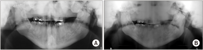
Osteopathia striata with cranial sclerosis (OS-CS) is a bone dysplasia characterized by a linear striated pattern of sclerosis, especially in the long bones, and cranial sclerosis. It has variable clinical findings but distinctive radiological findings. Multiple oral and dental findings have been associated with this disease and can be seen during dental and/or medical imaging of the head and neck. Dentists and clinicians must be familiar with these signs to differentiate them from pathosis or erroneous radiographs. In the following case, we present a patient with OS-CS that presented at The University of Florida College of Dentistry with multiple craniofacial manifestations of this syndrome that were seen on a panoramic radiograph, which is one of the most commonly requested radiographs by dentists.
条纹状骨病伴颅骨硬化(OS-CS)是一种骨发育异常,其特征为硬化的线性条纹状模式,尤其在长骨中,以及颅骨硬化。它有多种临床发现,但有独特的放射学表现。多种口腔和牙齿表现与该疾病相关,可在头颈部的牙科和/或医学影像检查中看到。牙医和临床医生必须熟悉这些体征,以便将它们与病变或错误的X光片区分开来。在以下病例中,我们介绍一名患有OS-CS的患者,该患者在佛罗里达大学牙科学院就诊,其全景X光片显示了该综合征的多种颅面表现,全景X光片是牙医最常要求拍摄的X光片之一。