Developmental Cognitive Neuroscience Lab (DCNL), Brain Institute, Pontifical Catholic University of Rio Grande do Sul (PUCRS), Porto Alegre, Brazil.
Louis A. Faillace, MD, Department of Psychiatry and Behavioral Sciences, University of Texas Health Science Center at Houston, Houston, TX, USA.
Transl Psychiatry. 2021 Apr 29;11(1):252. doi: 10.1038/s41398-021-01367-x.
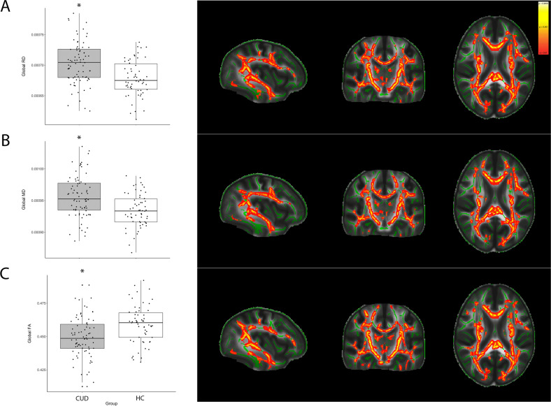
White matter (WM) abnormalities in patients with cocaine use disorder (CUD) have been studied; however, the reported effects on the human brain are heterogenous and most results have been obtained from male participants. In addition, biological data supporting the imaging findings and revealing possible mechanisms underlying the neurotoxic effects of chronic cocaine use (CU) on WM are largely restricted to animal studies. To evaluate the neurotoxic effects of CU in the WM, we performed an in vivo diffusion tensor imaging assessment of male and female cocaine users (n = 75) and healthy controls (HC) (n = 58). Moreover, we performed an ex vivo large-scale proteomic analysis using liquid chromatography-tandem mass spectrometry in postmortem brains of patients with CUD (n = 8) and HC (n = 12). Compared with the HC, the CUD group showed significant reductions in global fractional anisotropy (FA) (p < 0.001), and an increase in global mean (MD) and radial diffusion (RD) (both p < 0.001). The results revealed that FA, RD, and MD alterations in the CUD group were widespread along the major WM tracts, after analysis using the tract-based special statistics approach. Global FA was negatively associated with years of CU (p = 0.0421) and female sex (p < 0.001), but not with years of alcohol or nicotine use. Concerning the fibers connecting the left to the right prefrontal cortex, Brodmann area 9 (BA9), the CUD group presented lower FA (p = 0.006) and higher RD (p < 0.001) values compared with the HC group. A negative association between the duration of CU in life and FA values in this tract was also observed (p = 0.019). Proteomics analyses in BA9 found 11 proteins differentially expressed between cocaine users and controls. Among these, were proteins related to myelination and neuroinflammation. In summary, we demonstrate convergent evidence from in vivo diffusion tensor imaging and ex vivo proteomics analysis of WM disruption in CUD.
研究了可卡因使用障碍(CUD)患者的脑白质(WM)异常;然而,报告的对人类大脑的影响是异质的,并且大多数结果都是从男性参与者中获得的。此外,支持影像学发现并揭示慢性可卡因使用(CU)对 WM 神经毒性作用潜在机制的生物学数据在很大程度上仅限于动物研究。为了评估 CU 对 WM 的神经毒性作用,我们对 75 名男性和女性可卡因使用者(CUD)和 58 名健康对照(HC)进行了体内弥散张量成像评估。此外,我们使用液相色谱-串联质谱法对 8 名 CUD 患者和 12 名 HC 的死后大脑进行了大规模的蛋白质组学分析。与 HC 相比,CUD 组的整体分数各向异性(FA)显著降低(p < 0.001),整体平均(MD)和放射扩散(RD)增加(均 p < 0.001)。使用基于束的特殊统计学方法分析后,结果表明,CUD 组的 FA、RD 和 MD 改变广泛存在于主要 WM 束中。FA 与 CU 年限(p = 0.0421)和女性性别(p < 0.001)呈负相关,但与酒精或尼古丁使用年限无关。对于连接左右前额叶皮质的纤维,Brodmann 区 9(BA9),CUD 组的 FA 值较低(p = 0.006),RD 值较高(p < 0.001)与 HC 组相比。还观察到 CU 持续时间与该束中 FA 值之间的负相关(p = 0.019)。BA9 的蛋白质组学分析发现,可卡因使用者和对照组之间有 11 种蛋白质表达不同。其中包括与髓鞘形成和神经炎症相关的蛋白质。总之,我们从体内弥散张量成像和 WM 破坏的体外蛋白质组学分析中提供了一致的证据可卡因使用者。