Hakim Bilal Ahmad, Tyagi Vaishali, Agnihotri Saurabh Kumar, Nath Amar, Agrawal Ankit Kumar, Jain Ankita, Singh Deependra, Konwar Rituraj, Sachdev Monika
Division of Endocrinology, CSIR-Central Drug Research Institute (CDRI), Sector 10, Jankipuram Extension, Lucknow 226031, India.
Academy of Scientific and Innovative Research (AcSIR), New Delhi 110001, India.
J Dev Biol. 2021 Apr 1;9(2):13. doi: 10.3390/jdb9020013.
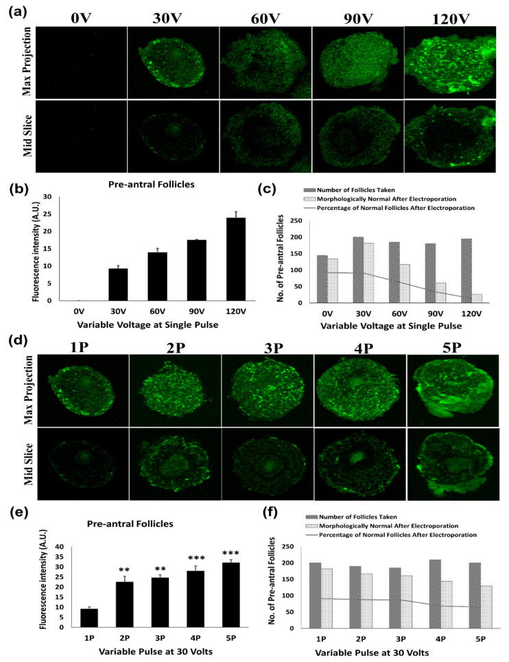
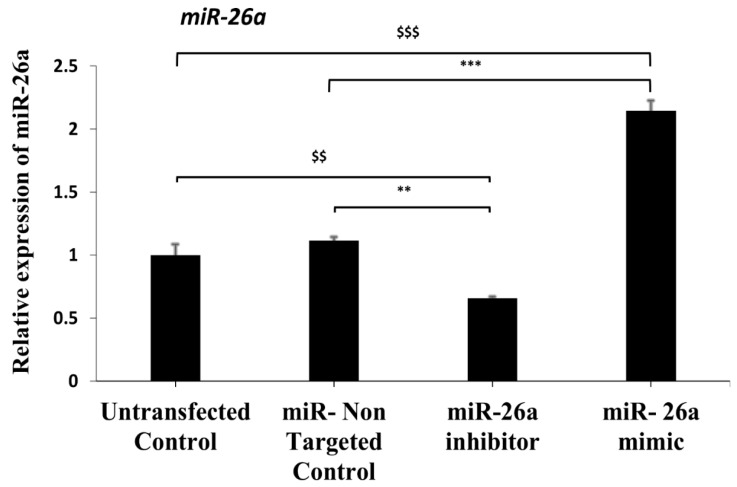
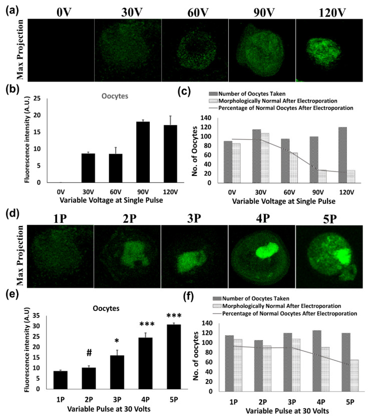
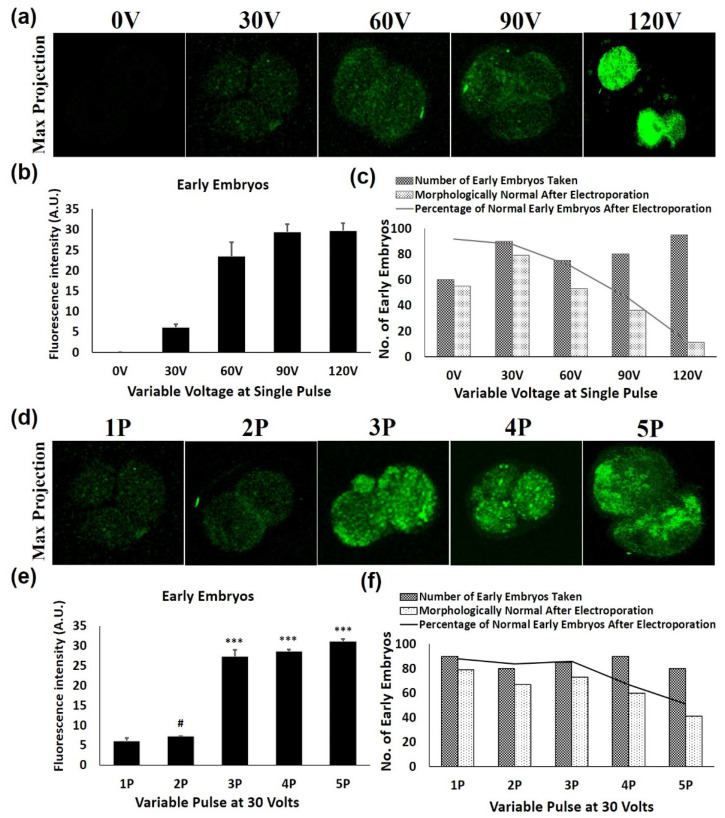
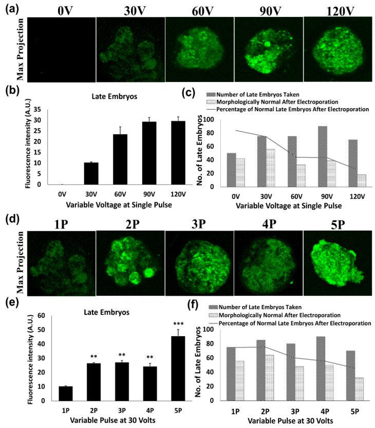
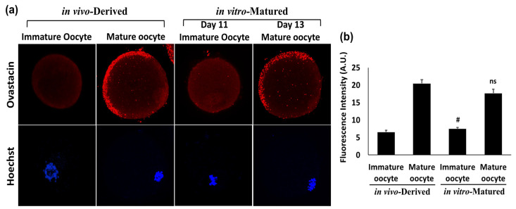
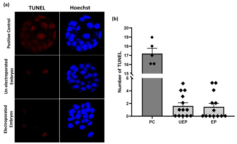
Electroporation is an effective technique of transfection, but its efficiency depends on the optimization of various parameters. In this study, a simplified and efficient method of gene manipulation was standardized through electroporation to introduce a recombinant green fluorescent protein (GFP) construct as well as RNA-inhibitors in intact mouse follicles, oocytes and early embryos, where various electroporation parameters like voltage, pulse number and pulse duration were standardized. Electroporated preantral follicles were cultured further in vitro to obtain mature oocytes and their viability was confirmed through the localization of a known oocyte maturation marker, ovastacin, which appeared to be similar to the in vivo-derived mature oocytes and thus proved the viability of the in vitro matured oocytes after electroporation. Standardized electroporation parameters, i.e., three pulses of 30 V for 1 millisecond at an interval of 10 s, were applied to manipulate the expression of mmu-miR-26a in preantral follicles through the electroporation of miR inhibitors and mimics. The TUNEL apoptosis assay confirmed the normal development of the electroporated embryos when compared to the normal embryos. Conclusively, for the first time, this study demonstrated the delivery of exogenous oligonucleotides into intact mouse follicles, oocytes and embryos without hampering their zona pellucida (ZP) and further development.
电穿孔是一种有效的转染技术,但其效率取决于各种参数的优化。在本研究中,通过电穿孔对一种简化高效的基因操作方法进行了标准化,以便将重组绿色荧光蛋白(GFP)构建体以及RNA抑制剂导入完整的小鼠卵泡、卵母细胞和早期胚胎中,同时对电压、脉冲数和脉冲持续时间等各种电穿孔参数进行了标准化。对电穿孔后的腔前卵泡进行进一步体外培养以获得成熟卵母细胞,并通过一种已知的卵母细胞成熟标志物卵母细胞溶素的定位来确认其活力,该标志物的定位似乎与体内来源的成熟卵母细胞相似,从而证明了电穿孔后体外成熟卵母细胞的活力。应用标准化的电穿孔参数,即30V的三个脉冲,持续1毫秒,间隔10秒,通过电穿孔导入miR抑制剂和模拟物来调控腔前卵泡中mmu-miR-26a的表达。TUNEL凋亡检测证实,与正常胚胎相比,电穿孔胚胎发育正常。总之,本研究首次证明了将外源寡核苷酸导入完整的小鼠卵泡、卵母细胞和胚胎中,而不会妨碍它们的透明带(ZP)和进一步发育。