Wähnert Dirk, Koettnitz Julian, Merten Madlen, Kronenberg Daniel, Stange Richard, Greiner Johannes F W, Kaltschmidt Christian, Vordemvenne Thomas, Kaltschmidt Barbara
Protestant Hospital of Bethel Foundation, Department of Trauma and Orthopedic Surgery, University Hospital OWL of Bielefeld University, Campus Bielefeld-Bethel, Burgsteig 13, 33617 Bielefeld, Germany.
Molecular Neurobiology, Bielefeld University, Universitätsstrasse 25, 33615 Bielefeld, Germany.
Materials (Basel). 2021 Apr 14;14(8):1961. doi: 10.3390/ma14081961.
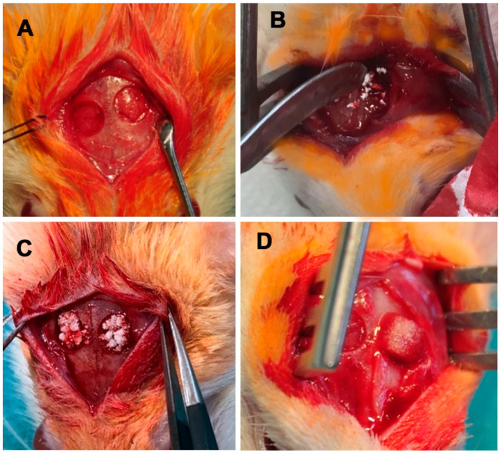
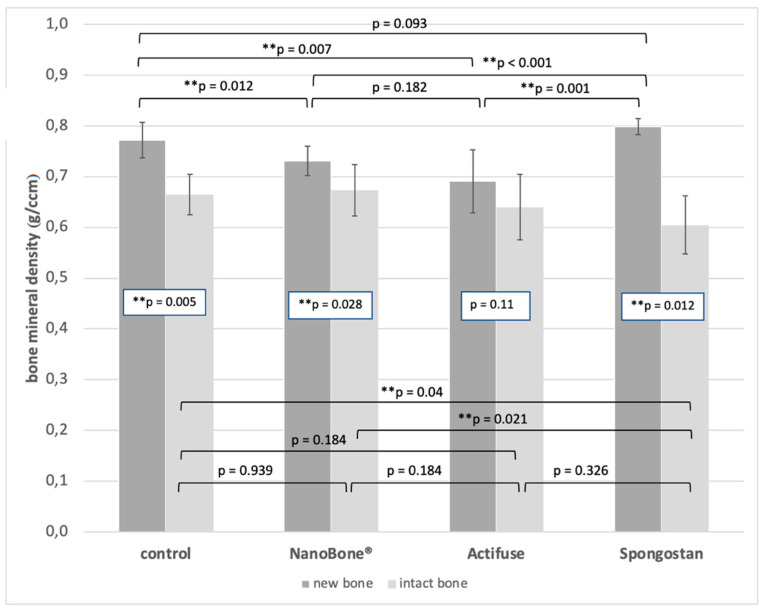
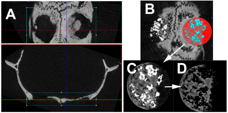
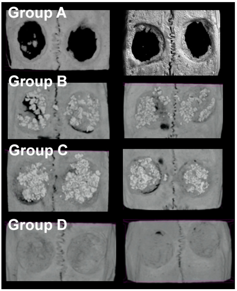
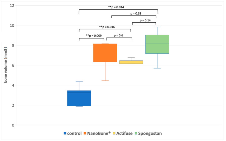
Bone substitute materials are becoming increasingly important in oral and maxillofacial surgery. Reconstruction of critical size bone defects is still challenging for surgeons. Here, we compared the clinically applied organic bone substitute materials NanoBone (nanocrystalline hydroxyapatite and nanostructured silica gel; = 5) and Actifuse (calcium phosphate with silicate substitution; = 5) with natural collagen-based Spongostan™ (hardened pork gelatin containing formalin and lauryl alcohol; = 5) in bilateral rat critical-size defects (5 mm diameter). On topological level, NanoBone is known to harbour nanopores of about 20 nm diameter, while Actifuse comprises micropores of 200-500 µm. Spongostan™, which is clinically applied as a haemostatic agent, combines in its wet form both nano- and microporous topological features by comprising 60.66 ± 24.48 μm micropores accompanied by nanopores of 32.97 ± 1.41 nm diameter. Micro-computed tomography (µCT) used for evaluation 30 days after surgery revealed a significant increase in bone volume by all three bone substitute materials in comparison to the untreated controls. Clearly visual was the closure of trepanation in all treated groups, but granular appearance of NanoBone and Actifuse with less closure at the margins of the burr holes. In contrast, transplantion of Spongostan™ lead to complete filling of the burr hole with the highest bone volume of 7.98 ccm and the highest bone mineral density compared to all other groups. In summary, transplantation of Spongostan™ resulted in increased regeneration of a rat calvarial critical size defect compared to NanoBone and Actifuse, suggesting the distinct nano- and microtopography of wet Spongostan™ to account for this superior regenerative capacity. Since Spongostan™ is a clinically approved product used primarily for haemostasis, it may represent an interesting alternative in the reconstruction of defects in the maxillary region.
骨替代材料在口腔颌面外科中正变得越来越重要。对于外科医生来说,关键尺寸骨缺损的重建仍然具有挑战性。在此,我们将临床应用的有机骨替代材料纳米骨(纳米晶羟基磷灰石和纳米结构硅胶;n = 5)和活性融合材料(含硅酸盐替代的磷酸钙;n = 5)与天然胶原蛋白基的海绵骨(含福尔马林和月桂醇的硬化猪明胶;n = 5)在双侧大鼠关键尺寸缺损(直径5毫米)中进行了比较。在拓扑层面,已知纳米骨含有直径约20纳米的纳米孔,而活性融合材料包含200 - 500微米的微孔。临床上用作止血剂的海绵骨,其湿态形式通过包含60.66 ± 24.48微米的微孔以及直径为32.97 ± 1.41纳米的纳米孔,兼具纳米和微孔拓扑特征。术后30天用于评估的微型计算机断层扫描(µCT)显示,与未治疗的对照组相比,所有三种骨替代材料的骨体积均显著增加。所有治疗组的钻孔闭合情况清晰可见,但纳米骨和活性融合材料呈颗粒状外观,钻孔边缘的闭合较少。相比之下,海绵骨的移植导致钻孔完全填充,骨体积最高达7.98立方厘米,且骨矿物质密度高于所有其他组。总之,与纳米骨和活性融合材料相比,海绵骨的移植导致大鼠颅骨关键尺寸缺损的再生增加,这表明湿态海绵骨独特的纳米和微观形貌是其具有这种卓越再生能力的原因。由于海绵骨是一种主要用于止血的临床批准产品,它可能是上颌区域缺损重建中一个有趣的替代选择。