Laboratory of Molecular Dietetics, Department of Neurological Diseases and Neurosurgery, Department of Analytical and Forensic Toxicology, IM Sechenov First Moscow State Medical University (Sechenov University), 119435 Moscow, Russia.
Laboratory of Ecobiomonitoring and Quality Control, Yaroslavl State University, 150003 Yaroslavl, Russia.
Int J Mol Sci. 2021 Apr 28;22(9):4646. doi: 10.3390/ijms22094646.
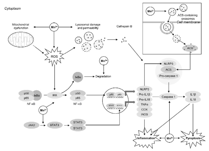
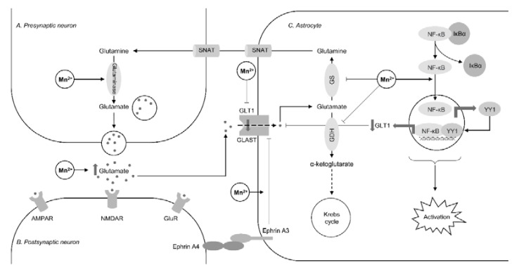
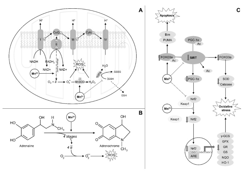
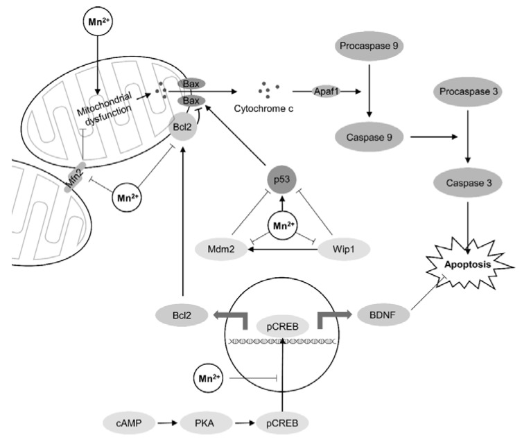
Understanding of the immediate mechanisms of Mn-induced neurotoxicity is rapidly evolving. We seek to provide a summary of recent findings in the field, with an emphasis to clarify existing gaps and future research directions. We provide, here, a brief review of pertinent discoveries related to Mn-induced neurotoxicity research from the last five years. Significant progress was achieved in understanding the role of Mn transporters, such as SLC39A14, SLC39A8, and SLC30A10, in the regulation of systemic and brain manganese handling. Genetic analysis identified multiple metabolic pathways that could be considered as Mn neurotoxicity targets, including oxidative stress, endoplasmic reticulum stress, apoptosis, neuroinflammation, cell signaling pathways, and interference with neurotransmitter metabolism, to name a few. Recent findings have also demonstrated the impact of Mn exposure on transcriptional regulation of these pathways. There is a significant role of autophagy as a protective mechanism against cytotoxic Mn neurotoxicity, yet also a role for Mn to induce autophagic flux itself and autophagic dysfunction under conditions of decreased Mn bioavailability. This ambivalent role may be at the crossroad of mitochondrial dysfunction, endoplasmic reticulum stress, and apoptosis. Yet very recent evidence suggests Mn can have toxic impacts below the no observed adverse effect of Mn-induced mitochondrial dysfunction. The impact of Mn exposure on supramolecular complexes SNARE and NLRP3 inflammasome greatly contributes to Mn-induced synaptic dysfunction and neuroinflammation, respectively. The aforementioned effects might be at least partially mediated by the impact of Mn on α-synuclein accumulation. In addition to Mn-induced synaptic dysfunction, impaired neurotransmission is shown to be mediated by the effects of Mn on neurotransmitter systems and their complex interplay. Although multiple novel mechanisms have been highlighted, additional studies are required to identify the critical targets of Mn-induced neurotoxicity.
对锰诱导神经毒性的即时机制的理解正在迅速发展。我们旨在提供该领域最新发现的综述,重点阐明现有差距和未来的研究方向。我们在此简要回顾了过去五年与锰诱导神经毒性研究相关的一些重要发现。在理解锰转运体(如 SLC39A14、SLC39A8 和 SLC30A10)在调节系统性和大脑锰处理中的作用方面取得了重大进展。遗传分析确定了多个代谢途径,这些途径可以被视为锰神经毒性的靶点,包括氧化应激、内质网应激、细胞凋亡、神经炎症、细胞信号通路以及干扰神经递质代谢等。最近的发现还表明,锰暴露对这些通路的转录调控有影响。自噬作为一种对抗细胞毒性锰神经毒性的保护机制具有重要作用,但锰也可以诱导自噬流本身,并在锰生物利用度降低的情况下导致自噬功能障碍。这种矛盾的作用可能处于线粒体功能障碍、内质网应激和细胞凋亡的交汇点。然而,最近的证据表明,在锰诱导线粒体功能障碍的无观察到不良效应水平以下,锰也可能产生毒性影响。锰暴露对 SNARE 和 NLRP3 炎性小体等超分子复合物的影响分别大大促进了锰诱导的突触功能障碍和神经炎症。上述影响至少部分可能是由于锰对α-突触核蛋白积累的影响所致。除了锰诱导的突触功能障碍外,还表明神经传递受损是由锰对神经递质系统及其复杂相互作用的影响介导的。尽管已经强调了多种新的机制,但还需要进行更多的研究来确定锰诱导神经毒性的关键靶点。