Suroto Heri, Aryawan Deny M, Prakoeswa Camilla A
Department of Orthopedics & Traumatology, Faculty of Medicine, Universitas Airlangga/Dr. Soetomo General Academic Hospital, Surabaya 60131, Indonesia.
Faculty of Medicine, Universitas Airlangga, Surabaya 60131, Indonesia.
Int J Biomater. 2021 Apr 7;2021:6685225. doi: 10.1155/2021/6685225. eCollection 2021.
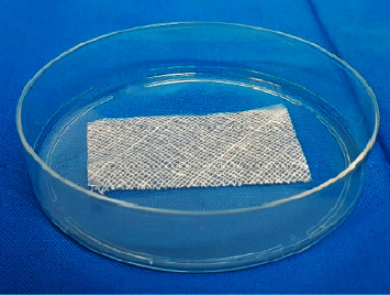
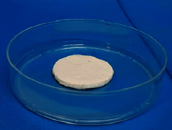
Amnion grafts can be preserved as freeze-dried amnion membrane (FD-AM) and amnion sponge. Preserved grafts require to be sterilized by gamma irradiation. However, each step of the process could affect its biological properties. Even so, there are only a few studies that report the influence of the preservation method and gamma irradiation on growth factor levels in preserved amniotic grafts.
This was an experimental study with a pretest-posttest group design using a consecutive sampling technique in one batch of amnion donors at a particular time. The amnion was made into FD-AM and amnion sponge preparations, and they were sterilized with gamma irradiation (15 kGy and 25 kGy). Nonirradiated specimens served as controls, and 20 mg of each specimen was pulverized to evaluate the growth factors levels using ELISA.
There were significant decreases in amnion sponge compared to the FD-AM, both in transforming growth factor beta (TGF-) and basic fibroblast growth factor (bFGF) levels and in the preirradiated and 25 kGy postirradiated preparations ( ≤ 0.05). The growth factor levels in the preirradiated and postirradiated FD-AM (both 15 kGy and 25 kGy) showed significant differences ( ≤ 0.05). Likewise, the preirradiated amnion sponge group's growth factor levels compared with the postirradiated amnion sponge group also showed a significant decrease ( ≤ 0.05).
TGF- and bFGF levels were lower in amnion sponge than FD-AM. The FD-AM and amnion sponge preparations' growth factors levels were reduced following gamma irradiation sterilization. Although the decrease in growth factor levels is significant, the number of growth factor levels is still sufficient for tissue healing.
羊膜移植物可制成冻干羊膜(FD - AM)和羊膜海绵。保存的移植物需要通过伽马射线照射进行灭菌。然而,该过程的每个步骤都可能影响其生物学特性。即便如此,仅有少数研究报告了保存方法和伽马射线照射对保存的羊膜移植物中生长因子水平的影响。
这是一项采用前后测试组设计的实验研究,在特定时间对一批羊膜供体采用连续抽样技术。将羊膜制成FD - AM和羊膜海绵制剂,并用伽马射线照射(15千戈瑞和25千戈瑞)进行灭菌。未照射的标本作为对照,将每个标本20毫克粉碎,用酶联免疫吸附测定法评估生长因子水平。
与FD - AM相比,羊膜海绵中的转化生长因子β(TGF - )和碱性成纤维细胞生长因子(bFGF)水平在照射前和25千戈瑞照射后的制剂中均显著降低(P≤0.05)。照射前和照射后(15千戈瑞和25千戈瑞)的FD - AM中的生长因子水平显示出显著差异(P≤0.05)。同样,照射前羊膜海绵组的生长因子水平与照射后羊膜海绵组相比也显著降低(P≤0.05)。
羊膜海绵中的TGF - 和bFGF水平低于FD - AM。伽马射线照射灭菌后,FD - AM和羊膜海绵制剂中的生长因子水平降低。尽管生长因子水平的降低显著,但生长因子的数量仍足以促进组织愈合。