Department of Radiology, Hainan General Hospital (Hainan Affiliated Hospital of Hainan Medical University), Haikou, China.
Department of Radiology, Weill Medical College of Cornell University, New York, NY, USA.
Cancer Sci. 2021 Jul;112(7):2835-2844. doi: 10.1111/cas.14918. Epub 2021 May 7.
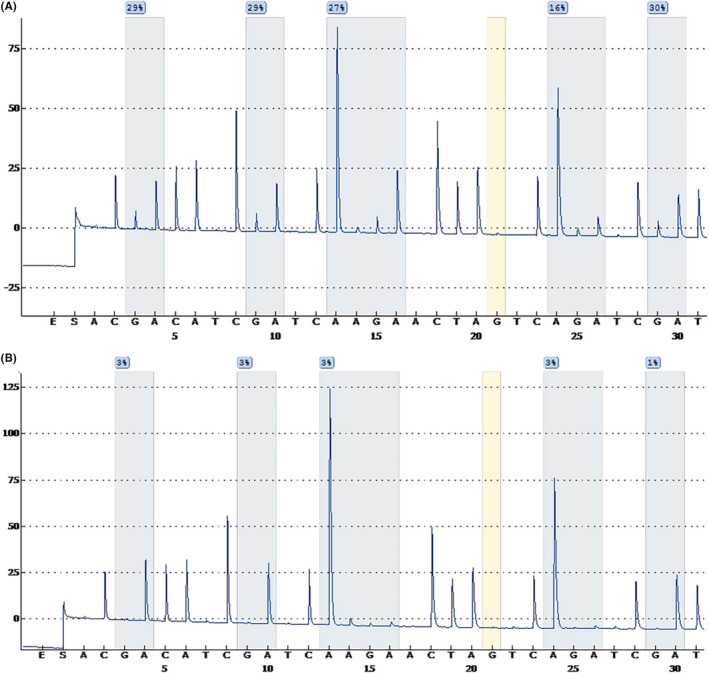
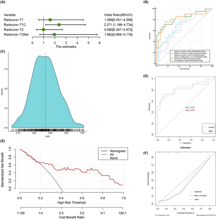
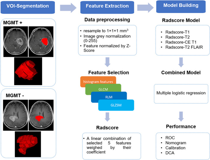
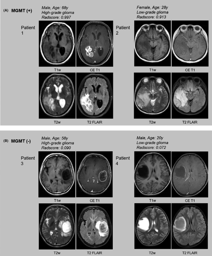
This study aims to build a radiological model based on standard MR sequences for detecting methylguanine methyltransferase (MGMT) methylation in gliomas using texture analysis. A retrospective cross-sectional study was undertaken in a cohort of 53 glioma patients who underwent standard preoperative magnetic resonance (MR) imaging. Conventional visual radiographic features and clinical factors were compared between MGMT promoter methylated and unmethylated groups. Texture analysis extracted the top five most powerful texture features of MR images in each sequence quantitatively for detecting the MGMT promoter methylation status. The radiomic signature (Radscore) was generated by a linear combination of the five features and estimates in each sequence. The combined model based on each Radscore was established using multivariate logistic regression analysis. A receiver operating characteristic (ROC) curve, nomogram, calibration, and decision curve analysis (DCA) were used to evaluate the performance of the model. No significant differences were observed in any of the visual radiographic features or clinical factors between different MGMT methylated statuses. The top five most powerful features were selected from a total of 396 texture features of T1, contrast-enhanced T1, T2, and T2 FLAIR. Each sequence's Radscore can distinguish MGMT methylated status. A combined model based on Radscores showed differentiation between methylated MGMT and unmethylated MGMT both in the glioblastoma (GBM) dataset as well as the dataset for all other gliomas. The area under the ROC curve values for the combined model was 0.818, with 90.5% sensitivity and 72.7% specificity, in the GBM dataset, and 0.833, with 70.2% sensitivity and 90.6% specificity, in the overall gliomas dataset. Nomogram, calibration, and DCA also validated the performance of the combined model. The combined model based on texture features could be considered as a noninvasive imaging marker for detecting MGMT methylation status in glioma.
本研究旨在建立一种基于标准磁共振(MR)序列的放射影像学模型,通过纹理分析来检测脑胶质瘤中甲基鸟嘌呤甲基转移酶(MGMT)的甲基化状态。该研究为回顾性病例对照研究,纳入了 53 例接受标准术前磁共振成像检查的脑胶质瘤患者。比较了 MGMT 启动子甲基化和非甲基化组之间的常规影像学特征和临床因素。通过定量分析每个序列的磁共振图像,提取了前 5 个最具特征性的纹理特征,用于检测 MGMT 启动子的甲基化状态。放射组特征(Radscore)由 5 个特征的线性组合和每个序列中的估计值生成。基于每个 Radscore 的多变量逻辑回归分析建立了综合模型。使用受试者工作特征(ROC)曲线、列线图、校准和决策曲线分析(DCA)评估模型的性能。不同 MGMT 甲基化状态之间的任何视觉影像学特征或临床因素均无显著差异。从 T1、增强 T1、T2 和 T2 FLAIR 共 396 个纹理特征中筛选出前 5 个最具特征性的特征。每个序列的 Radscore 都可以区分 MGMT 甲基化状态。基于 Radscore 的综合模型可区分脑胶质瘤(GBM)数据集和所有其他脑胶质瘤数据集的甲基化 MGMT 和非甲基化 MGMT。在 GBM 数据集中,综合模型的 ROC 曲线下面积值为 0.818,灵敏度为 90.5%,特异性为 72.7%;在所有脑胶质瘤数据集中,ROC 曲线下面积值为 0.833,灵敏度为 70.2%,特异性为 90.6%。列线图、校准和 DCA 也验证了综合模型的性能。基于纹理特征的综合模型可作为脑胶质瘤 MGMT 甲基化状态的一种非侵入性影像学标志物。