• 文献检索
  • 文档翻译
  • 深度研究
  • 学术资讯
  • Suppr Zotero 插件Zotero 插件
  • 邀请有礼
  • 套餐&价格
  • 历史记录
应用&插件
Suppr Zotero 插件Zotero 插件浏览器插件Mac 客户端Windows 客户端微信小程序
定价
高级版会员购买积分包购买API积分包
服务
文献检索文档翻译深度研究API 文档MCP 服务
关于我们
关于 Suppr公司介绍联系我们用户协议隐私条款
关注我们

Suppr 超能文献

核心技术专利:CN118964589B侵权必究
粤ICP备2023148730 号-1Suppr @ 2026

文献检索

告别复杂PubMed语法,用中文像聊天一样搜索,搜遍4000万医学文献。AI智能推荐,让科研检索更轻松。

立即免费搜索

文件翻译

保留排版,准确专业,支持PDF/Word/PPT等文件格式,支持 12+语言互译。

免费翻译文档

深度研究

AI帮你快速写综述,25分钟生成高质量综述,智能提取关键信息,辅助科研写作。

立即免费体验

印度东北部日本脑炎的磁共振波谱:一项横断面研究。

MRI Spectrum of Japanese Encephalitis in Northeast India: A Cross-Sectional Study.

作者信息

Phukan Pranjal, Sarma Kalyan, Sharma Barun Kumar, Boruah Deb K, Gogoi Bidyut Bikash, Chuunthang Daniala

机构信息

Department of Radiodiagnosis, North Eastern Indira Gandhi Regional Institute of Health and Medical Sciences, Shillong, Meghalaya, India.

Department of Neuroradiology, All India Institute of Medical Sciences, New Delhi, India.

出版信息

J Neurosci Rural Pract. 2021 Apr;12(2):281-289. doi: 10.1055/s-0041-1722820. Epub 2021 Mar 24.

DOI:10.1055/s-0041-1722820
PMID:33935446
原文链接:https://pmc.ncbi.nlm.nih.gov/articles/PMC8079177/
Abstract

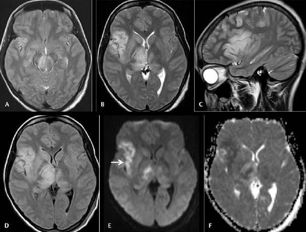
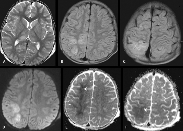
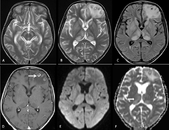
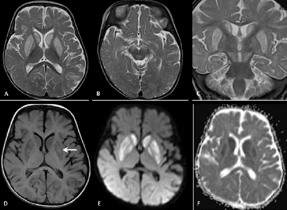
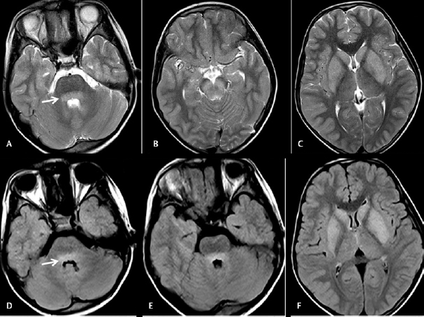
Japanese encephalitis (JE) is an arthropod-borne flavivirus infection having high mortality and morbidity. This study was performed to evaluate the conventional magnetic resonance imaging (MRI) findings in JE and to find out any difference between pediatric and adult JE.  This retrospective study was performed on serologically positive 54 JE patients presented to a tertiary care hospital with acute encephalitic symptoms between April 2016 and October 2019. Relevant neurological examination, cerebrospinal fluid analysis, and MRI scan of the brain were performed.  Fifty-four JE patients ( = 31 males and = 23 females) having 32 pediatric and 22 adult JE were included in the study sample. Group 1 JE ( = 16) patients had encephalitic symptoms with duration less than 15 days up to the day of MRI scan and group 2 JE ( = 38) had symptoms more than 15 days. Group 1 JE had mean apparent diffusion coefficient (ADC) value of 0.563 ± 0.109 (standard deviation [SD]) × 10 mm /sec and group 2 JE had 1.095 ± 0.206 (SD) × 10 mm /sec. The mean ADC value of pediatric JE was 0.907 ± 0.336 (SD) × 10 mm /sec and adult JE was 0.982 ± 0.253 (SD) × 10 mm /sec.  The majority of the JE patient shows abnormal signal alterations in bilateral thalami and substantia nigra. Diffusion-weighted imaging with ADC mapping helps in evaluating the stage of the JE. No statistical significance of the various conventional MRI findings was found between the pediatric JE and adult JE.

摘要

日本脑炎(JE)是一种由节肢动物传播的黄病毒感染,具有较高的死亡率和发病率。本研究旨在评估JE患者的传统磁共振成像(MRI)表现,并找出小儿和成人JE之间的差异。

本回顾性研究纳入了2016年4月至2019年10月期间在一家三级医院就诊的54例血清学阳性的JE患者,这些患者均出现急性脑炎症状。对患者进行了相关的神经学检查、脑脊液分析和脑部MRI扫描。

研究样本包括54例JE患者(男性31例,女性22例),其中小儿JE患者32例,成人JE患者22例。第1组JE患者(16例)在MRI扫描当天出现脑炎症状的持续时间少于15天,第2组JE患者(38例)症状持续时间超过15天。第1组JE患者的平均表观扩散系数(ADC)值为0.563±0.109(标准差[SD])×10⁻³mm²/sec,第2组JE患者为1.095±0.206(SD)×10⁻³mm²/sec。小儿JE患者的平均ADC值为0.907±0.336(SD)×10⁻³mm²/sec,成人JE患者为0.982±0.253(SD)×10⁻³mm²/sec。

大多数JE患者在双侧丘脑和黑质出现异常信号改变。弥散加权成像及ADC图有助于评估JE的阶段。小儿JE和成人JE在各种传统MRI表现上未发现统计学差异。

https://cdn.ncbi.nlm.nih.gov/pmc/blobs/0d37/8079177/dcbe190d1863/10-1055-s-0041-1722820_08_1045_05.jpg
https://cdn.ncbi.nlm.nih.gov/pmc/blobs/0d37/8079177/52c016b54ce5/10-1055-s-0041-1722820_08_1045_01.jpg
https://cdn.ncbi.nlm.nih.gov/pmc/blobs/0d37/8079177/917db6974cb2/10-1055-s-0041-1722820_08_1045_02.jpg
https://cdn.ncbi.nlm.nih.gov/pmc/blobs/0d37/8079177/10ac3671dfbe/10-1055-s-0041-1722820_08_1045_03.jpg
https://cdn.ncbi.nlm.nih.gov/pmc/blobs/0d37/8079177/42697fbba0c1/10-1055-s-0041-1722820_08_1045_04.jpg
https://cdn.ncbi.nlm.nih.gov/pmc/blobs/0d37/8079177/dcbe190d1863/10-1055-s-0041-1722820_08_1045_05.jpg

相似文献

1
MRI Spectrum of Japanese Encephalitis in Northeast India: A Cross-Sectional Study.印度东北部日本脑炎的磁共振波谱:一项横断面研究。
J Neurosci Rural Pract. 2021 Apr;12(2):281-289. doi: 10.1055/s-0041-1722820. Epub 2021 Mar 24.
2
Diffusion-weighted imaging and apparent diffusion coefficient evaluation of herpes simplex encephalitis and Japanese encephalitis.单纯疱疹病毒性脑炎和日本脑炎的弥散加权成像及表观弥散系数评估。
J Neurol Sci. 2009 Dec 15;287(1-2):221-6. doi: 10.1016/j.jns.2009.07.010. Epub 2009 Sep 3.
3
A Profile Study of Japanese Encephalitis in an Industrial Hospital in Eastern India.印度东部一家工业医院的日本脑炎概况研究。
Cureus. 2023 May 2;15(5):e38455. doi: 10.7759/cureus.38455. eCollection 2023 May.
4
Diffusion-weighted MR imaging in Japanese encephalitis.
J Comput Assist Tomogr. 2004 Nov-Dec;28(6):756-61. doi: 10.1097/00004728-200411000-00005.
5
Diffusion-weighted MR imaging of viral encephalitis.病毒性脑炎的磁共振扩散加权成像
Neuroradiology. 2006 Dec;48(12):875-80. doi: 10.1007/s00234-006-0143-7. Epub 2006 Aug 31.
6
[A case of Japanese encephalitis presenting with unilateral lesions in diffusion-weighted MRI].1例在扩散加权磁共振成像中表现为单侧病变的日本脑炎病例
Rinsho Shinkeigaku. 2001 Sep;41(9):602-5.
7
An appraisal of clinicopathological parameters in Japanese encephalitis and changing epidemiological trends in upper Assam, India.印度上阿萨姆邦日本脑炎的临床病理参数评估及流行病学趋势变化
Indian J Pathol Microbiol. 2014 Jul-Sep;57(3):400-6. doi: 10.4103/0377-4929.138732.
8
Clinical, radiological and neurophysiological spectrum of JEV encephalitis and other non-specific encephalitis during post-monsoon period in India.印度季风后期日本脑炎病毒脑炎及其他非特异性脑炎的临床、放射学和神经生理学特征
Neurol India. 2003 Mar;51(1):55-9.
9
Clinical and radiological spectrum of Japanese encephalitis.日本脑炎的临床和放射学特征。
J Neurol Sci. 2013 Feb 15;325(1-2):15-21. doi: 10.1016/j.jns.2012.11.007. Epub 2012 Dec 20.
10
A comparison of clinical features of Japanese encephalitis virus infection in the adult and pediatric age group with Acute Encephalitis Syndrome.成人和儿童年龄组的日本脑炎病毒感染与急性脑炎综合征的临床特征比较。
J Clin Virol. 2011 Sep;52(1):45-9. doi: 10.1016/j.jcv.2011.06.001. Epub 2011 Jun 28.

引用本文的文献

1
The alarming spread of Japanese encephalitis: A growing public health concern.日本脑炎的惊人传播:日益严重的公共卫生问题。
Open Vet J. 2025 Apr;15(4):1505-1519. doi: 10.5455/OVJ.2025.v15.i4.1. Epub 2025 Apr 30.
2
An Atypical Presentation of Posterior Reversible Encephalopathy Syndrome (PRES): Sticking to the Basics.后部可逆性脑病综合征(PRES)的非典型表现:回归基础
Cureus. 2024 Oct 19;16(10):e71836. doi: 10.7759/cureus.71836. eCollection 2024 Oct.
3
Neuroinvasion of emerging and re-emerging arboviruses: A scoping review.

本文引用的文献

1
Japanese Encephalitis presenting with cerebral venous sinus thrombosis: a case report.
Med J Malaysia. 2019 Dec;74(6):537-539.
2
Longitudinal magnetic resonance imaging changes in Japanese encephalitis.日本脑炎的纵向磁共振成像变化
Brain Dev. 2019 Sep;41(8):731-734. doi: 10.1016/j.braindev.2019.04.005. Epub 2019 Apr 16.
3
Japanese encephalitis.日本脑炎
CMAJ. 2018 May 28;190(21):E657. doi: 10.1503/cmaj.171341.
新发和再发虫媒病毒的神经侵袭:一项范围综述
SAGE Open Med. 2024 May 6;12:20503121241229847. doi: 10.1177/20503121241229847. eCollection 2024.
4
Clinical characteristics and short-term outcomes of Japanese encephalitis in pediatric and adult patients: a retrospective study in Northern China.中国北方儿童和成人日本脑炎的临床特征及短期预后:一项回顾性研究
Front Neurol. 2023 Apr 20;14:1135001. doi: 10.3389/fneur.2023.1135001. eCollection 2023.
5
Japanese Encephalitis among Adults: A Review.成人日本脑炎:综述。
Am J Trop Med Hyg. 2023 Apr 10;108(5):860-864. doi: 10.4269/ajtmh.23-0036. Print 2023 May 3.
6
Who Is at Risk? A Critical Case of Japanese Encephalitis.谁处于危险之中?一例日本脑炎的重症病例。
J Acute Med. 2022 Sep 1;12(3):122-125. doi: 10.6705/j.jacme.202209_12(3).0005.
4
Diagnostic dilemma-dengue or Japanese encephalitis?诊断难题——登革热还是日本脑炎?
Neurol India. 2017 Jan-Feb;65(1):105-107. doi: 10.4103/0028-3886.198194.
5
Bilateral thalamic bleed and cerebral venous sinus thrombosis in Japanese encephalitis.日本脑炎中的双侧丘脑出血和脑静脉窦血栓形成
BMJ Case Rep. 2015 Jan 7;2015:bcr2014207957. doi: 10.1136/bcr-2014-207957.
6
Japanese encephalitis: the virus and vaccines.日本脑炎:病毒与疫苗
Hum Vaccin Immunother. 2014;10(2):263-79. doi: 10.4161/hv.26902. Epub 2013 Oct 25.
7
Limbic encephalitis: a clinical-radiological comparison between herpetic and autoimmune etiologies.边缘性脑炎:疱疹性和自身免疫性病因的临床-放射学比较。
Eur J Neurol. 2013 Dec;20(12):1566-70. doi: 10.1111/ene.12249. Epub 2013 Aug 14.
8
Clinical and radiological spectrum of Japanese encephalitis.日本脑炎的临床和放射学特征。
J Neurol Sci. 2013 Feb 15;325(1-2):15-21. doi: 10.1016/j.jns.2012.11.007. Epub 2012 Dec 20.
9
MRI features of Japanese encephalitis.日本脑炎的磁共振成像特征。
BMJ Case Rep. 2012 Jul 3;2012:bcr0320126088. doi: 10.1136/bcr.03.2012.6088.
10
Japanese encephalitis accompanied by cerebral venous sinus thrombosis: a case report.日本脑炎合并脑静脉窦血栓形成:一例报告。
BMC Neurol. 2012 Jun 19;12:43. doi: 10.1186/1471-2377-12-43.