Li Jing, Zhang Qihao, Che Yena, Zhang Nan, Guo Lingfei
Department of Radiology, Beijing Friendship Hospital, Capital Medical University, Beijing, China.
Department of Radiology, Weill Cornell Medical College, Cornell University, New York City, NY, United States.
Front Aging Neurosci. 2021 Apr 14;13:611891. doi: 10.3389/fnagi.2021.611891. eCollection 2021.
The objective of this study was to determine which factors influence brain iron concentrations in deep gray matter in elderly individuals and how these factors influence regional brain iron concentrations.
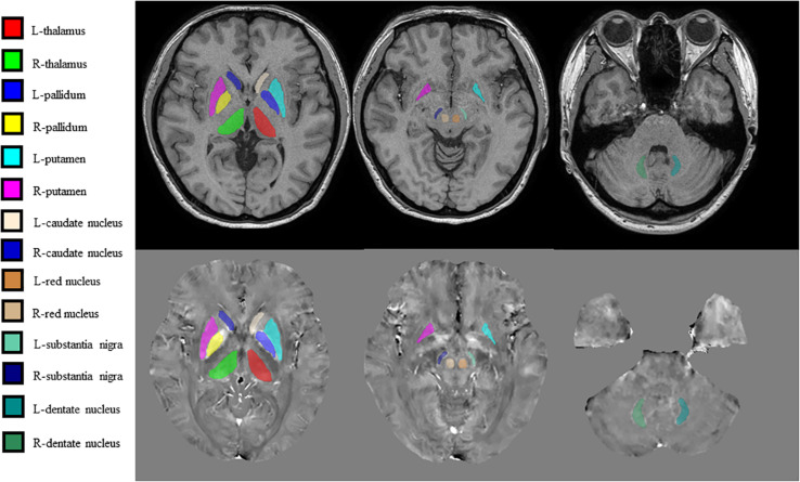
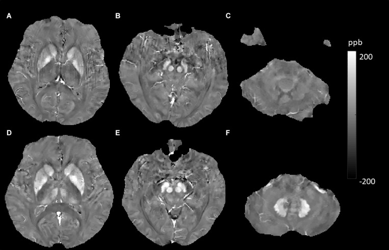
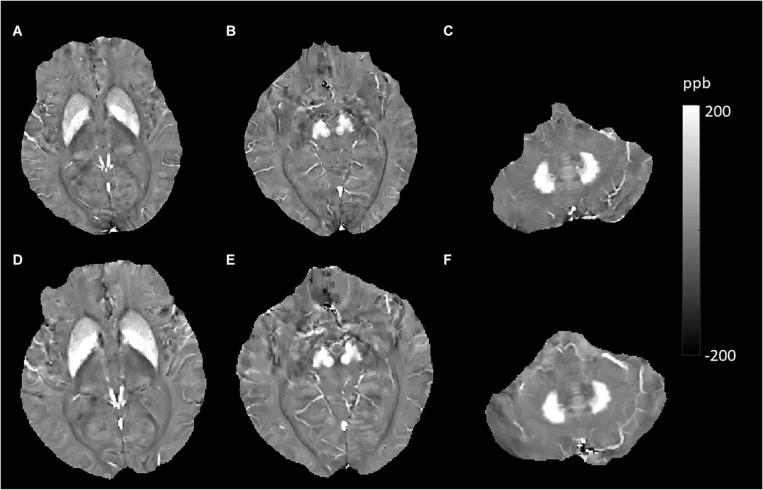
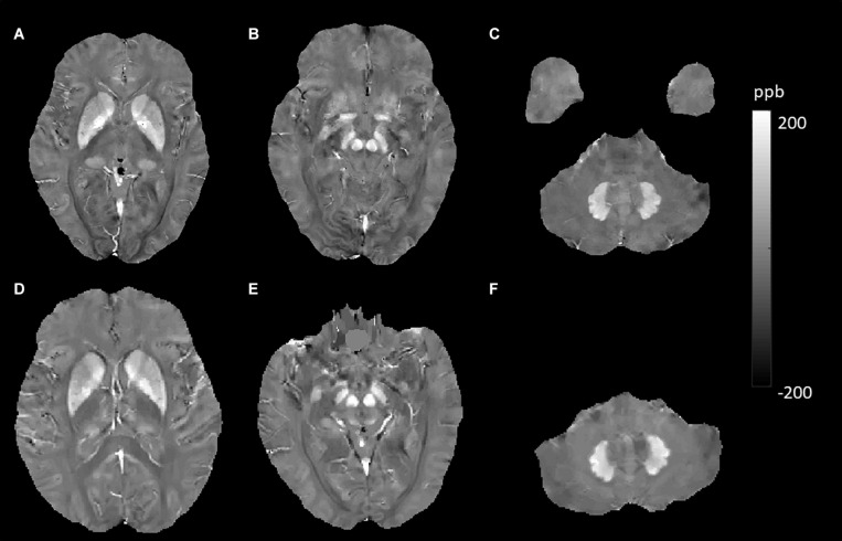
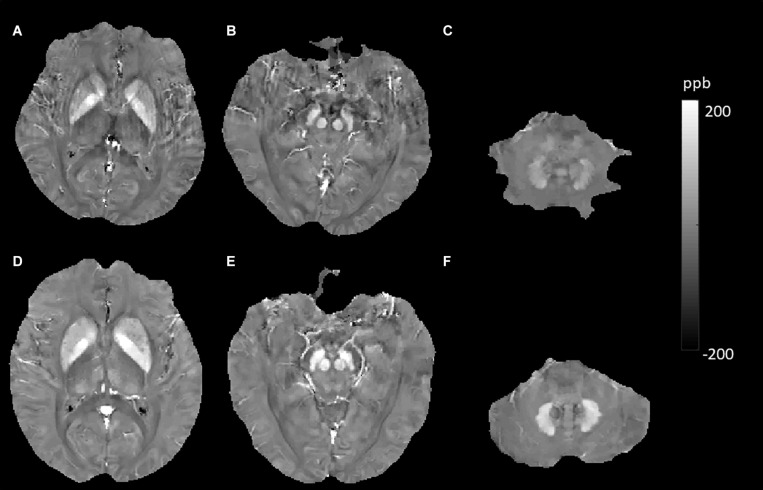
A total of 105 elderly individuals were enrolled in this study. All participants underwent detailed magnetic resonance imaging (MRI) examinations from October 2018 to August 2019. Among them, 44 individuals had undergone a previous MRI examination from July 2010 to August 2011. Quantitative susceptibility mapping (QSM) was utilized as an indirect quantitative marker of brain iron, and the susceptibility values of deep gray matter structures were obtained. Univariate analysis and multiple linear regression analysis were used to investigate 11 possible determinants for cerebral iron deposition.
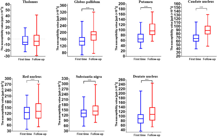
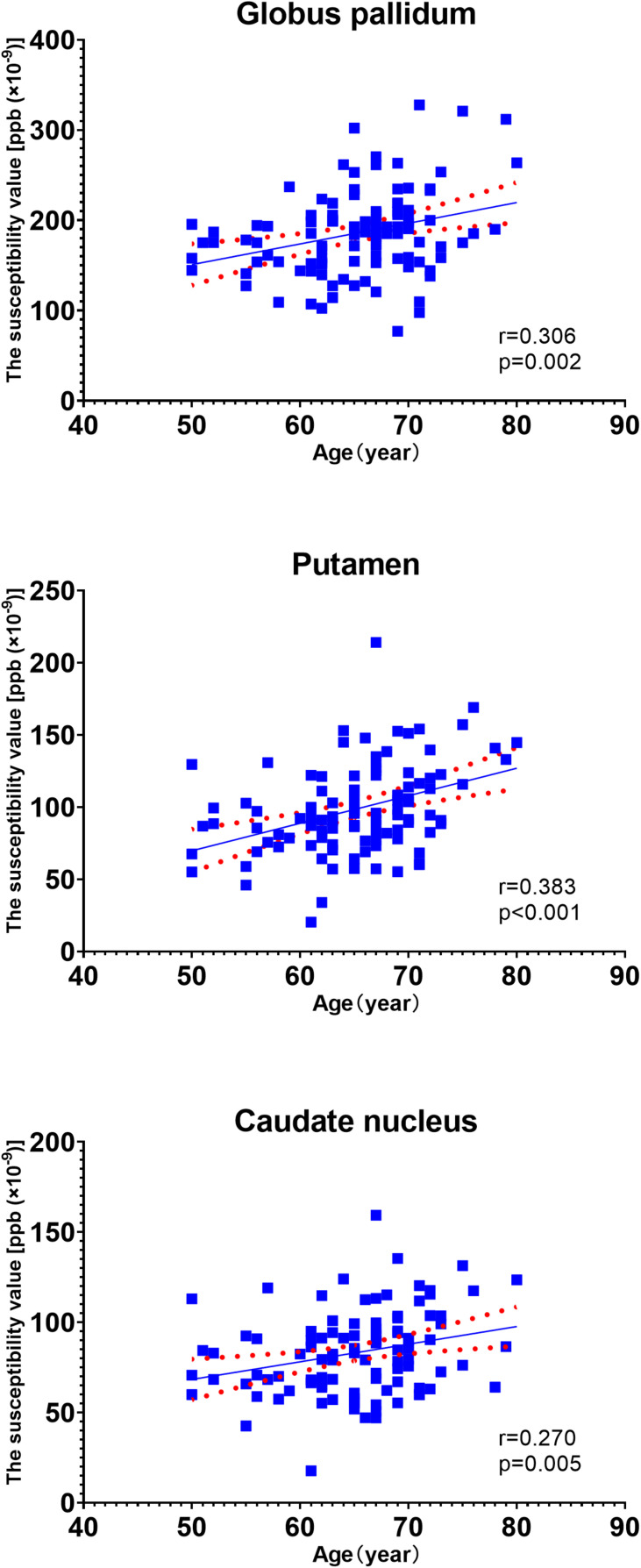
Our results showed no sex- or hemisphere-related differences in susceptibility values in any of the regions studied. Aging was significantly correlated with increased insusceptibility values in almost all analyzed brain regions (except for the thalamus) when we compared the susceptibility values at the two time points. In a cross-sectional analysis, the relationship between gray matter nucleus susceptibility values and age was conducted using Pearson's linear regression. Aging was significantly correlated with the susceptibility values of the globus pallidus (GP), putamen (Put), and caudate nucleus (CN), with the Put having the strongest correlations. In multiple linear regression models, associations with increased susceptibility values were found in the CN, Put, red nucleus, and dentate nucleus for individuals with a history of type 2 diabetes mellitus (T2DM). However, the patients with hypertension showed significantly reduced susceptibility values in the red nucleus and dentate nucleus. Our data suggested that smokers had increased susceptibility values in the thalamus. No significant associations were found for individuals with a history of hypercholesterolemia and Apolipoprotein E4 carrier status.
Our data revealed that aging, T2DM, and smoking could increase iron deposition in some deep gray matter structures. However, hypertension had the opposite effects in the red nuclei and dentate nuclei. Brain iron metabolism could be influenced by many factors in different modes. In future studies, we should strictly control for confounding factors.
本研究的目的是确定哪些因素影响老年人深部灰质中的脑铁浓度,以及这些因素如何影响局部脑铁浓度。
本研究共纳入105名老年人。所有参与者在2018年10月至2019年8月期间接受了详细的磁共振成像(MRI)检查。其中,44人在2010年7月至2011年8月期间曾接受过MRI检查。定量磁化率成像(QSM)被用作脑铁的间接定量指标,并获取深部灰质结构的磁化率值。采用单因素分析和多元线性回归分析来研究脑铁沉积的11个可能决定因素。
我们的结果显示,在所研究的任何区域中,磁化率值均无性别或半球相关差异。当我们比较两个时间点的磁化率值时,衰老与几乎所有分析脑区(除丘脑外)的磁化率增加显著相关。在横断面分析中,使用Pearson线性回归分析灰质核磁化率值与年龄之间的关系。衰老与苍白球(GP)、壳核(Put)和尾状核(CN)的磁化率值显著相关,其中壳核的相关性最强。在多元线性回归模型中,2型糖尿病(T2DM)病史患者的CN、Put、红核和齿状核的磁化率值增加。然而,高血压患者的红核和齿状核磁化率值显著降低。我们的数据表明,吸烟者丘脑的磁化率值增加。高胆固醇血症病史和载脂蛋白E4携带者状态的个体未发现显著关联。
我们的数据显示,衰老、T2DM和吸烟可增加某些深部灰质结构中的铁沉积。然而,高血压在红核和齿状核中有相反的作用。脑铁代谢可能受多种因素以不同方式影响。在未来的研究中,我们应严格控制混杂因素。