Department of Periodontics, Texas A&M University, College of Dentistry, Dallas, Texas, USA.
Department of Biomedical Sciences, Texas A&M University, College of Dentistry, Dallas, Texas, USA.
Clin Exp Dent Res. 2021 Oct;7(5):679-691. doi: 10.1002/cre2.412. Epub 2021 May 3.
Many acellular dermal matrices (ADMs) are available for use in periodontal surgical procedures. However, few studies exist evaluating their in vivo healing properties. The objectives of this study were to compare the wound healing and remodeling of two ADMs used for gingival augmentation procedures in the rat model.
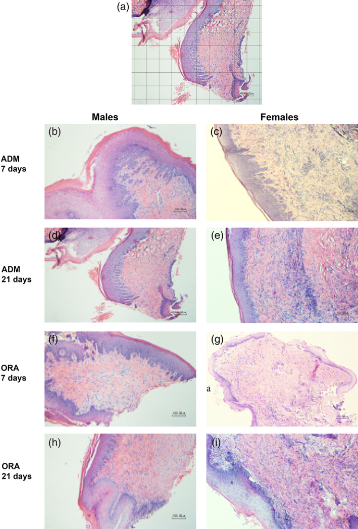
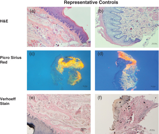
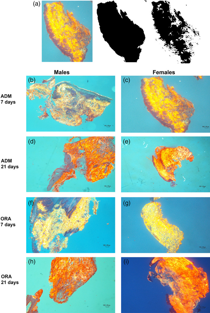
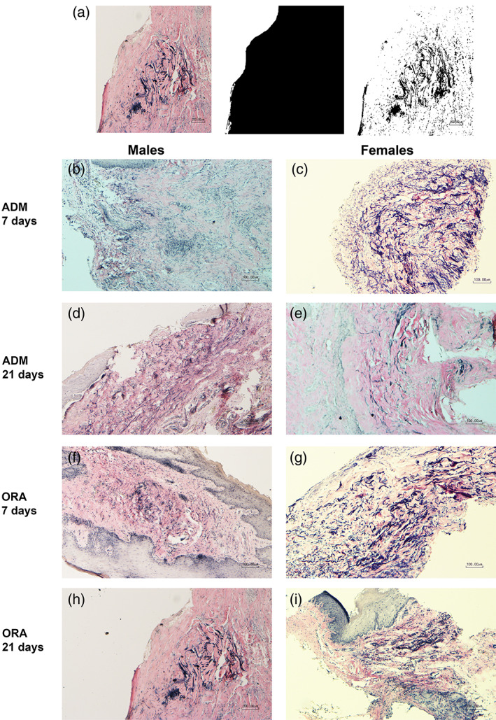
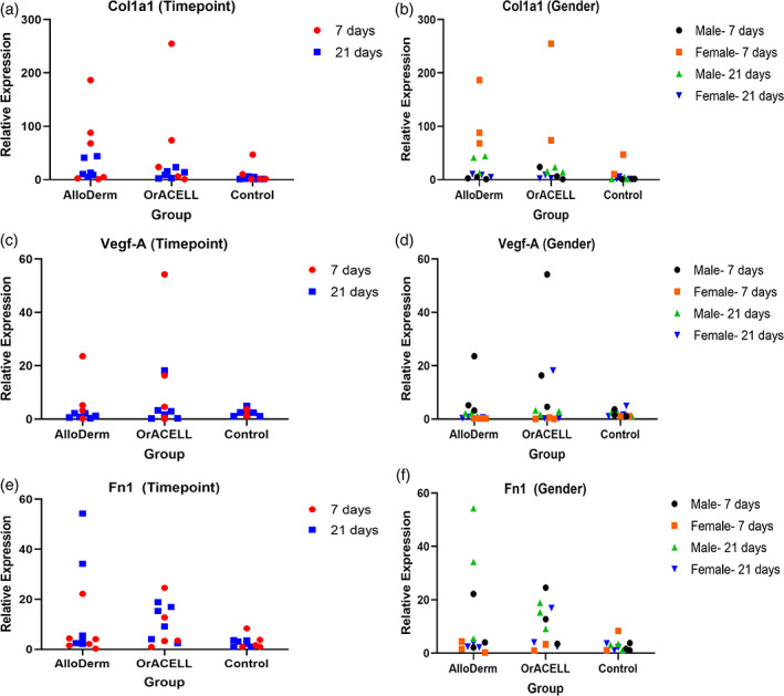
This was a nonrandomized controlled split-mouth study. Envelope flaps were surgically created in the maxillary quadrants of 24 Sprague Dawley rats. Each received either (a) AlloDerm Regenerative Tissue Matrix, or (b) OrACELL. Gingival tissue from one mandibular quadrant served as the untreated control. Six male and six female rats were treated for 7 or 21 days. Biopsies were processed for histologic analysis (H&E, Picro-sirius red, Verhoeff's solution) or RNA analysis (RT-PCR) to analyze the expression of type I collagen (Col1a1), fibronectin (Fn-1) and VEGF-A (Vegf-A).
There was a greater density of fibroblasts in OrACELL compared to AlloDerm at both timepoints. There was a greater density of elastin present in AlloDerm compared to OrACELL at 7 days but no differences at 21 days. There were no differences between test groups in the percentage of birefringent collagen or in the expression of Vegf-A or Fn-1. At 7 days, there were significantly more fibroblasts for males in the OrACELL group compared to females. At 21 days, there was a significantly greater expression of Col1a1 for males in the OrACELL group compared to females.
Early wound healing and remodeling of OrACELL appeared to occur more rapidly than AlloDerm and was accelerated in male rats. Whether these results have clinical implications for soft tissue grafting procedures in humans remains to be determined.
有许多脱细胞真皮基质(ADM)可用于牙周手术。然而,目前评估其体内愈合特性的研究很少。本研究的目的是比较两种用于牙龈增大术的 ADM 在大鼠模型中的伤口愈合和重塑。
这是一项非随机对照的分侧研究。在 24 只 Sprague Dawley 大鼠的上颌象限中手术创建信封瓣。每只大鼠接受以下两种治疗之一:(a)AlloDerm 再生组织基质,或(b)OrACELL。一个下颌象限的牙龈组织作为未处理的对照。6 只雄性和 6 只雌性大鼠分别治疗 7 天或 21 天。对活检标本进行组织学分析(H&E、Picro-sirius 红、Verhoeff 溶液)或 RNA 分析(RT-PCR),以分析 I 型胶原(Col1a1)、纤维连接蛋白(Fn-1)和 VEGF-A(Vegf-A)的表达。
在两个时间点,OrACELL 中的成纤维细胞密度均高于 AlloDerm。在 7 天时,AlloDerm 中的弹性蛋白密度高于 OrACELL,但在 21 天时没有差异。在测试组中,双折射胶原的百分比、Vegf-A 或 Fn-1 的表达均无差异。在 7 天时,OrACELL 组雄性大鼠的成纤维细胞密度明显高于雌性大鼠。在 21 天时,OrACELL 组雄性大鼠的 Col1a1 表达明显高于雌性大鼠。
OrACELL 的早期伤口愈合和重塑似乎比 AlloDerm 更快,并且在雄性大鼠中更快。这些结果是否对人类软组织移植手术有临床意义仍有待确定。