Department of Radiology, Yamaguchi University Graduate School of Medicine, 1-1-1 Minamikogushi, Ube, Yamaguchi, 755-8505, Japan.
Department of Radiology, National Hospital Organization Yamaguchi - Ube Medical Center, 685 Higashikiwa, Ube, Yamaguchi, 755-0241, Japan.
Jpn J Radiol. 2021 Sep;39(9):868-876. doi: 10.1007/s11604-021-01122-8. Epub 2021 May 4.
The purpose of this study was to compare the high-resolution CT (HRCT) findings of pulmonary infectious and noninfectious complications with extensive ground-glass attenuation (GGA) in immunocompromised patients.
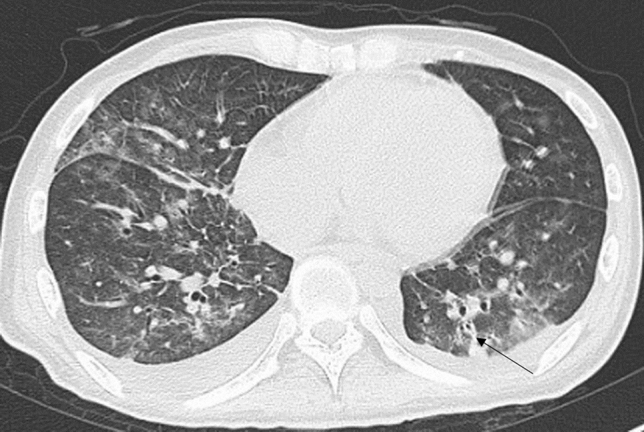
One hundred fifty-two immunocompromised patients with pulmonary complications that showed extensive GGA (> 50% of the whole lung on HRCT) were included in this study. The diagnoses of the 152 patients were as follows: pneumocystis pneumonia (PCP), n = 82; drug-induced pneumonia, n = 38; bacterial pneumonia, n = 9; cytomegalovirus pneumonia, n = 6; idiopathic pneumonia syndrome, n = 6; diffuse alveolar hemorrhage (DAH), n = 4; fungal infection, n = 3; tuberculosis, n = 2 and pulmonary edema, n = 2. Two chest radiologists retrospectively evaluated the CT criteria, which consisted of 12 findings.
The nodule (p = 0.015), the bronchovascular bundle (BVB) thickening (p = 0.001), and the interlobular septum (ILS) thickening (p = 0.002) were significantly infrequent in PCP. The ILS thickening was significantly frequent in drug-induced pneumonia (p < 0.001) though it was also frequent in other noninfectious and infectious diseases. The BVB thickening was significantly frequent in bacterial pneumonia (p = 0.005). The nodule was significantly frequent in DAH (p = 0.049).
Nodules, BVB thickening, and ILS thickening could be useful HRCT findings for the differential diagnosis of pulmonary complications in immunocompromised patients with extensive GGA.
本研究旨在比较免疫功能低下患者广泛磨玻璃影(GGA)肺部感染性和非感染性并发症的高分辨率 CT(HRCT)表现。
本研究纳入了 152 例 GGA 广泛(HRCT 上>50%的整个肺)的免疫功能低下肺部并发症患者。152 例患者的诊断如下:卡氏肺孢子虫肺炎(PCP),n=82;药物性肺炎,n=38;细菌性肺炎,n=9;巨细胞病毒肺炎,n=6;特发性肺炎综合征,n=6;弥漫性肺泡出血(DAH),n=4;真菌感染,n=3;肺结核,n=2 和肺水肿,n=2。两位胸部放射科医生回顾性评估了 CT 标准,该标准包括 12 种表现。
结节(p=0.015)、支气管血管束(BVB)增厚(p=0.001)和小叶间隔(ILS)增厚(p=0.002)在 PCP 中明显不常见。尽管 ILS 增厚在其他非感染性和感染性疾病中也很常见,但在药物性肺炎中更为常见(p<0.001)。BVB 增厚在细菌性肺炎中更为常见(p=0.005)。结节在 DAH 中更为常见(p=0.049)。
结节、BVB 增厚和 ILS 增厚可能是鉴别免疫功能低下患者广泛 GGA 肺部并发症的有用 HRCT 表现。