The Catholic University Liver Research Center, Department of Biomedicine & Health Sciences, College of Medicine, The Catholic University of Korea, Seoul 06591, Korea.
Department of Internal Medicine, College of Medicine, Seoul St. Mary's Hospital, The Catholic University of Korea, Seoul 06591, Korea.
Int J Mol Sci. 2021 Apr 29;22(9):4710. doi: 10.3390/ijms22094710.
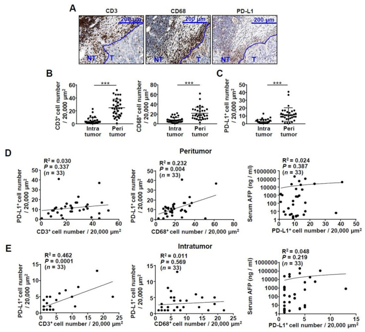
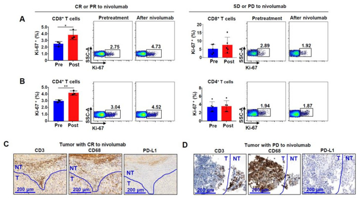
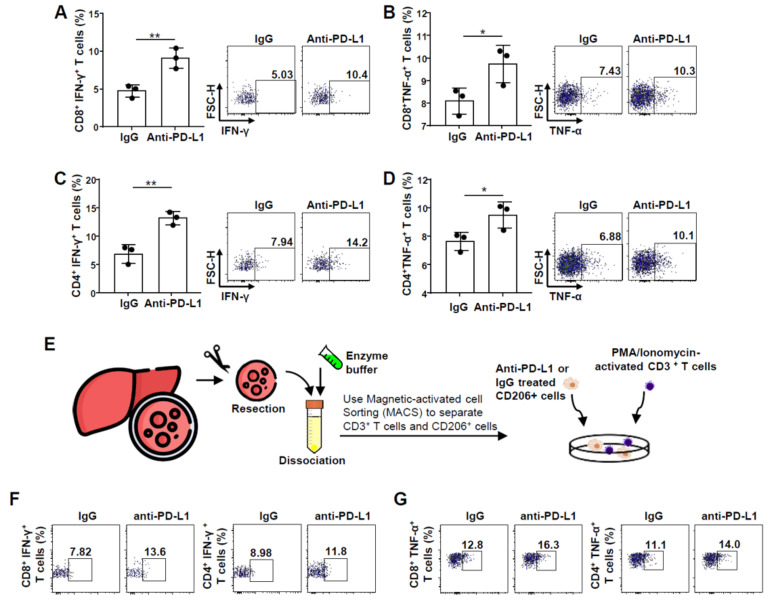
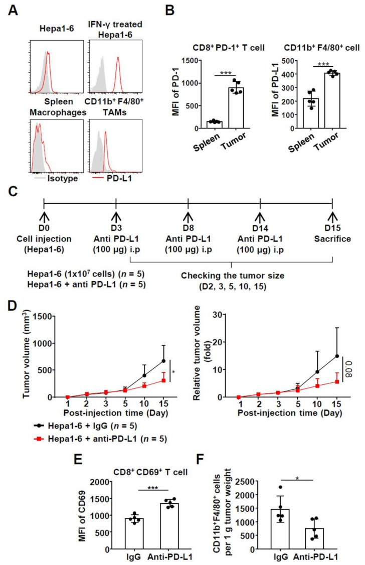
A predictive biomarker of immune checkpoint inhibitor (ICI)-based treatments in hepatocellular carcinoma (HCC) has not been clearly demonstrated. In this study, we focused on the infiltration and programmed death ligand 1 (PD-L1) expression of tumor-associated macrophages (TAMs) in the tumor microenvironment of HCC. Immunohistochemistry demonstrated that PD-L1 was preferentially expressed on CD68 macrophages in the tumor microenvironment of HCC, suggestive of its expression in TAMs rather than in T cells or tumor cells ( < 0.05). A co-culture experiment using activated T cells and M2 macrophages confirmed a significant increase in T cell functionality after the pretreatment of M2 macrophages with anti-PD-L1. Syngeneic mouse model experiments demonstrated that TAMs expressed PD-L1 and tumors treated with anti-PD-L1 showed smaller diameters than those treated with IgG. In these mice, anti-PD-L1 treatment increased activation markers in intratumoral CD8 T cells and reduced the size of the TAM population. Regarding nivolumab-treated patients, three of eight patients responded to the anti-PD-1 treatment. The percentage of Ki-67-positive CD4 and CD8 T cells was higher in responders than non-responders after nivolumab. Overall, PD-L1 expression on TAMs may be targeted by immune-based HCC treatment, and ICI treatment results in the reinvigoration of exhausted CD8 T cells in HCC.
在肝细胞癌 (HCC) 中,尚未明确预测免疫检查点抑制剂 (ICI) 治疗的生物标志物。在这项研究中,我们专注于 HCC 肿瘤微环境中肿瘤相关巨噬细胞 (TAM) 的浸润和程序性死亡配体 1 (PD-L1) 表达。免疫组织化学显示,PD-L1 在 HCC 肿瘤微环境中的 CD68 巨噬细胞中优先表达,提示其在 TAM 中表达,而不是在 T 细胞或肿瘤细胞中表达(<0.05)。使用激活的 T 细胞和 M2 巨噬细胞进行的共培养实验证实,在用抗 PD-L1 预处理 M2 巨噬细胞后,T 细胞功能显著增强。同基因小鼠模型实验表明 TAMs 表达 PD-L1,用抗 PD-L1 治疗的肿瘤直径小于用 IgG 治疗的肿瘤。在这些小鼠中,抗 PD-L1 治疗增加了肿瘤内 CD8 T 细胞中的激活标志物,并减少了 TAM 群体的大小。关于纳武利尤单抗治疗的患者,8 名患者中有 3 名对抗 PD-1 治疗有反应。与无反应者相比,纳武利尤单抗治疗后,Ki-67 阳性 CD4 和 CD8 T 细胞的百分比在应答者中更高。总体而言,TAM 上的 PD-L1 表达可能成为基于免疫的 HCC 治疗的靶点,ICI 治疗可使 HCC 中耗竭的 CD8 T 细胞恢复活力。