• 文献检索
  • 文档翻译
  • 深度研究
  • 学术资讯
  • Suppr Zotero 插件Zotero 插件
  • 邀请有礼
  • 套餐&价格
  • 历史记录
应用&插件
Suppr Zotero 插件Zotero 插件浏览器插件Mac 客户端Windows 客户端微信小程序
定价
高级版会员购买积分包购买API积分包
服务
文献检索文档翻译深度研究API 文档MCP 服务
关于我们
关于 Suppr公司介绍联系我们用户协议隐私条款
关注我们

Suppr 超能文献

核心技术专利:CN118964589B侵权必究
粤ICP备2023148730 号-1Suppr @ 2026

文献检索

告别复杂PubMed语法,用中文像聊天一样搜索,搜遍4000万医学文献。AI智能推荐,让科研检索更轻松。

立即免费搜索

文件翻译

保留排版,准确专业,支持PDF/Word/PPT等文件格式,支持 12+语言互译。

免费翻译文档

深度研究

AI帮你快速写综述,25分钟生成高质量综述,智能提取关键信息,辅助科研写作。

立即免费体验

用于钻石光源长波长大分子晶体学光束线的X射线断层扫描重建与分割流程

X-ray tomographic reconstruction and segmentation pipeline for the long-wavelength macromolecular crystallography beamline at Diamond Light Source.

作者信息

Kazantsev Daniil, Duman Ramona, Wagner Armin, Mykhaylyk Vitaliy, Wanelik Kazimir, Basham Mark, Wadeson Nicola

机构信息

Diamond Light Source Ltd, Diamond House, Harwell Science and Innovation Campus, Fermi Avenue, Didcot OX11 0DE United Kingdom.

出版信息

J Synchrotron Radiat. 2021 May 1;28(Pt 3):889-901. doi: 10.1107/S1600577521003453. Epub 2021 Apr 20.

DOI:10.1107/S1600577521003453
PMID:33949996
原文链接:https://pmc.ncbi.nlm.nih.gov/articles/PMC8127374/
Abstract

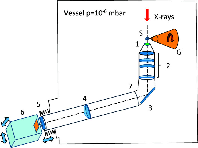
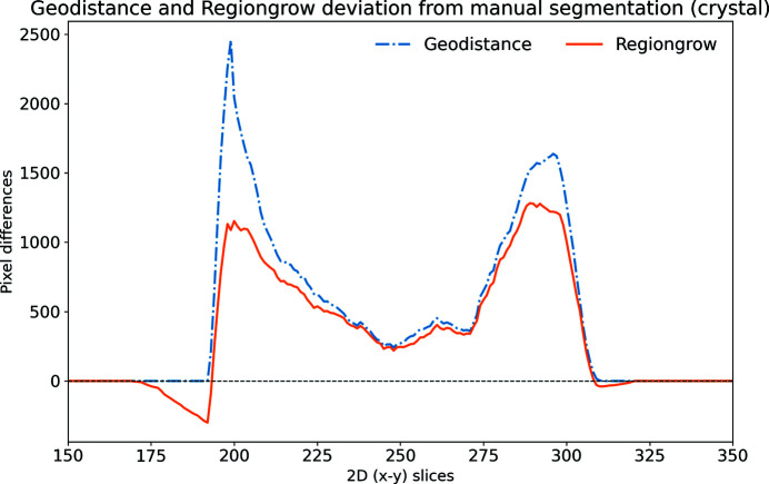
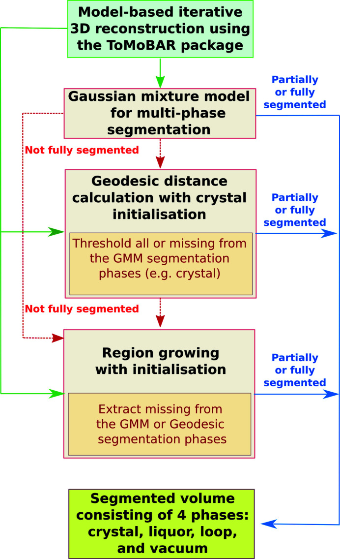
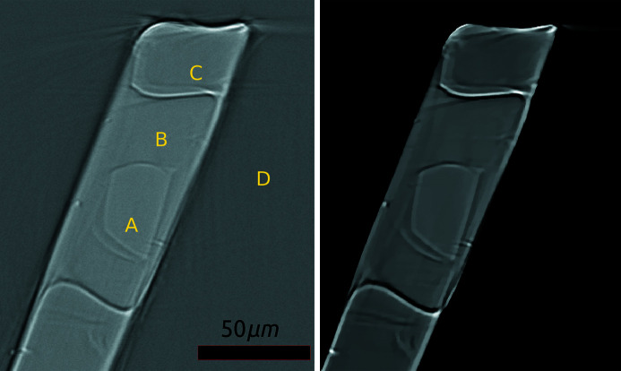
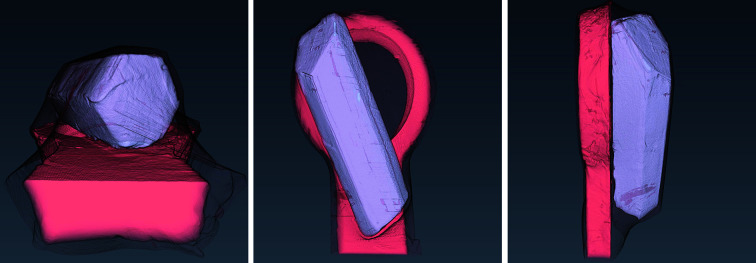
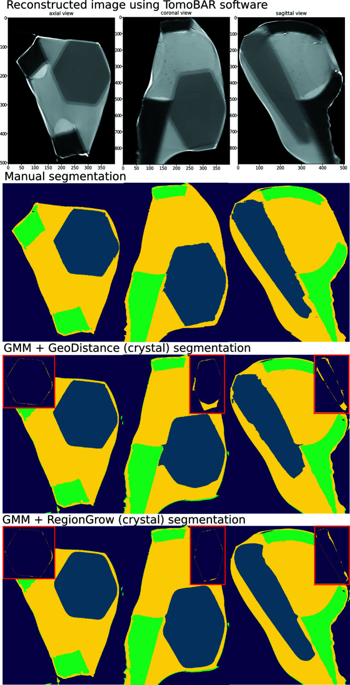
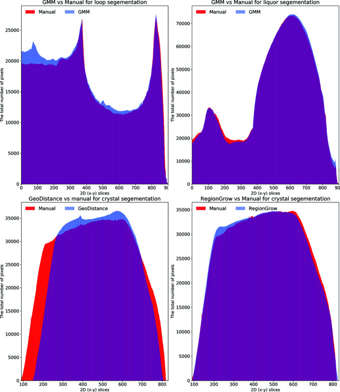
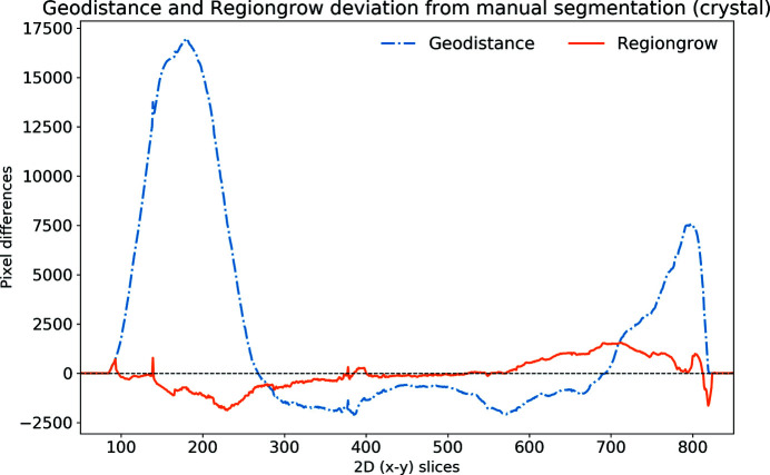
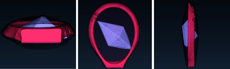
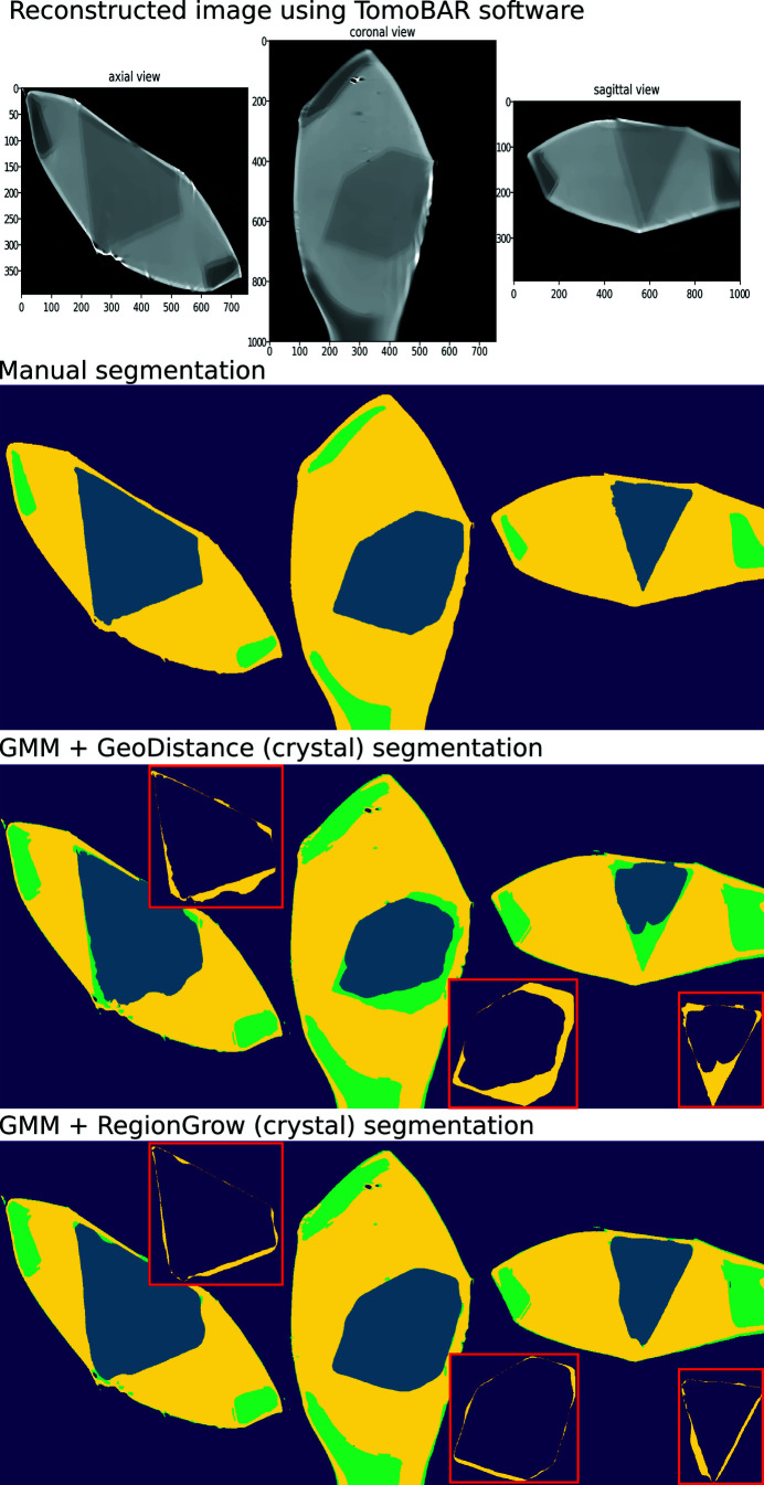
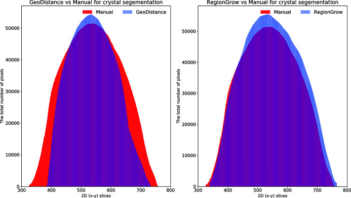
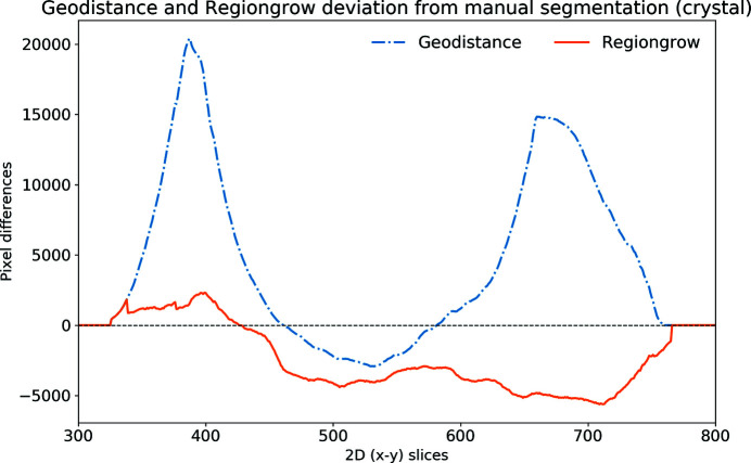
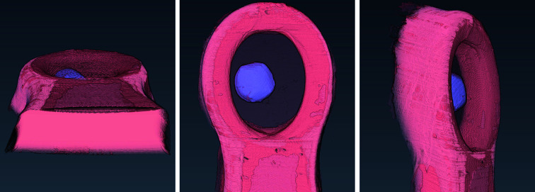
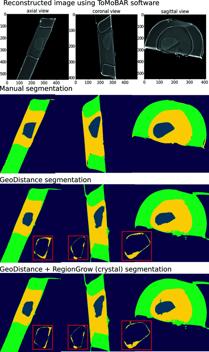
In this paper a practical solution for the reconstruction and segmentation of low-contrast X-ray tomographic data of protein crystals from the long-wavelength macromolecular crystallography beamline I23 at Diamond Light Source is provided. The resulting segmented data will provide the path lengths through both diffracting and non-diffracting materials as basis for analytical absorption corrections for X-ray diffraction data taken in the same sample environment ahead of the tomography experiment. X-ray tomography data from protein crystals can be difficult to analyse due to very low or absent contrast between the different materials: the crystal, the sample holder and the surrounding mother liquor. The proposed data processing pipeline consists of two major sequential operations: model-based iterative reconstruction to improve contrast and minimize the influence of noise and artefacts, followed by segmentation. The segmentation aims to partition the reconstructed data into four phases: the crystal, mother liquor, loop and vacuum. In this study three different semi-automated segmentation methods are experimented with by using Gaussian mixture models, geodesic distance thresholding and a novel morphological method, RegionGrow, implemented specifically for the task. The complete reconstruction-segmentation pipeline is integrated into the MPI-based data analysis and reconstruction framework Savu, which is used to reduce computation time through parallelization across a computing cluster and makes the developed methods easily accessible.

摘要

本文提供了一种实用的解决方案,用于对来自钻石光源长波长大分子晶体学光束线I23的蛋白质晶体低对比度X射线断层扫描数据进行重建和分割。所得的分割数据将提供穿过衍射和非衍射材料的路径长度,作为在断层扫描实验之前在相同样品环境中采集的X射线衍射数据的分析吸收校正的基础。由于不同材料(晶体、样品架和周围的母液)之间的对比度非常低或不存在,蛋白质晶体的X射线断层扫描数据可能难以分析。所提出的数据处理流程包括两个主要的顺序操作:基于模型的迭代重建,以提高对比度并最小化噪声和伪影的影响,随后进行分割。分割旨在将重建数据划分为四个阶段:晶体、母液、环和真空。在本研究中,通过使用高斯混合模型、测地距离阈值化和专门为该任务实现的一种新颖的形态学方法RegionGrow,对三种不同的半自动分割方法进行了试验。完整的重建 - 分割流程被集成到基于MPI的数据分析和重建框架Savu中,该框架用于通过跨计算集群并行化来减少计算时间,并使所开发的方法易于使用。

https://cdn.ncbi.nlm.nih.gov/pmc/blobs/73dd/8127374/b5ed60fa0b80/s-28-00889-fig17.jpg
https://cdn.ncbi.nlm.nih.gov/pmc/blobs/73dd/8127374/3cfca9f90749/s-28-00889-fig1.jpg
https://cdn.ncbi.nlm.nih.gov/pmc/blobs/73dd/8127374/585e48ad605c/s-28-00889-fig2.jpg
https://cdn.ncbi.nlm.nih.gov/pmc/blobs/73dd/8127374/155ec45bbf6f/s-28-00889-fig3.jpg
https://cdn.ncbi.nlm.nih.gov/pmc/blobs/73dd/8127374/dfda553226ad/s-28-00889-fig4.jpg
https://cdn.ncbi.nlm.nih.gov/pmc/blobs/73dd/8127374/2e82ac0adb27/s-28-00889-fig5.jpg
https://cdn.ncbi.nlm.nih.gov/pmc/blobs/73dd/8127374/ac39c23fccab/s-28-00889-fig6.jpg
https://cdn.ncbi.nlm.nih.gov/pmc/blobs/73dd/8127374/ee05ba4d6eb7/s-28-00889-fig7.jpg
https://cdn.ncbi.nlm.nih.gov/pmc/blobs/73dd/8127374/f68a94e53b5a/s-28-00889-fig8.jpg
https://cdn.ncbi.nlm.nih.gov/pmc/blobs/73dd/8127374/204b36ca4b67/s-28-00889-fig9.jpg
https://cdn.ncbi.nlm.nih.gov/pmc/blobs/73dd/8127374/c3d1b66b7b3d/s-28-00889-fig10.jpg
https://cdn.ncbi.nlm.nih.gov/pmc/blobs/73dd/8127374/13ec68fa3816/s-28-00889-fig11.jpg
https://cdn.ncbi.nlm.nih.gov/pmc/blobs/73dd/8127374/89c723caa1a5/s-28-00889-fig12.jpg
https://cdn.ncbi.nlm.nih.gov/pmc/blobs/73dd/8127374/424cd328294d/s-28-00889-fig13.jpg
https://cdn.ncbi.nlm.nih.gov/pmc/blobs/73dd/8127374/3d6c58e305f6/s-28-00889-fig14.jpg
https://cdn.ncbi.nlm.nih.gov/pmc/blobs/73dd/8127374/9ee0945e3ff6/s-28-00889-fig15.jpg
https://cdn.ncbi.nlm.nih.gov/pmc/blobs/73dd/8127374/8fe5307a1d0b/s-28-00889-fig16.jpg
https://cdn.ncbi.nlm.nih.gov/pmc/blobs/73dd/8127374/b5ed60fa0b80/s-28-00889-fig17.jpg

相似文献

1
X-ray tomographic reconstruction and segmentation pipeline for the long-wavelength macromolecular crystallography beamline at Diamond Light Source.用于钻石光源长波长大分子晶体学光束线的X射线断层扫描重建与分割流程
J Synchrotron Radiat. 2021 May 1;28(Pt 3):889-901. doi: 10.1107/S1600577521003453. Epub 2021 Apr 20.
2
A Sample Preparation Pipeline for Microcrystals at the VMXm Beamline.VMXm 光束线上的微晶体样品制备流水线。
J Vis Exp. 2021 Jun 17(172). doi: 10.3791/62306.
3
Sample Preparation and Transfer Protocol for In-Vacuum Long-Wavelength Crystallography on Beamline I23 at Diamond Light Source.真空长波长晶体学在钻石光源 I23 光束线上的样品制备和传输协议。
J Vis Exp. 2021 Apr 23(170). doi: 10.3791/62364.
4
Structure of the cyanobactin oxidase ThcOx from Cyanothece sp. PCC 7425, the first structure to be solved at Diamond Light Source beamline I23 by means of S-SAD.来自集胞藻 PCC 7425 的蓝细菌素氧化酶 ThcOx 的结构,这是首个在 Diamond Light Source 光束线 I23 通过 S-SAD 方法解析的结构。
Acta Crystallogr D Struct Biol. 2016 Nov 1;72(Pt 11):1174-1180. doi: 10.1107/S2059798316015850. Epub 2016 Oct 28.
5
Ray-tracing analytical absorption correction for X-ray crystallography based on tomographic reconstructions.基于断层重建的X射线晶体学光线追踪解析吸收校正
J Appl Crystallogr. 2024 Apr 15;57(Pt 3):649-658. doi: 10.1107/S1600576724002243. eCollection 2024 Jun 1.
6
Long-wavelength macromolecular crystallography - First successful native SAD experiment close to the sulfur edge.长波长大分子晶体学——首次在硫边缘附近成功进行的天然单波长反常散射实验。
Nucl Instrum Methods Phys Res B. 2017 Nov 15;411:12-16. doi: 10.1016/j.nimb.2016.12.005.
7
In-vacuum long-wavelength macromolecular crystallography.真空内长波长大分子晶体学
Acta Crystallogr D Struct Biol. 2016 Mar;72(Pt 3):430-9. doi: 10.1107/S2059798316001078. Epub 2016 Mar 1.
8
A high-throughput system for high-quality tomographic reconstruction of large datasets at Diamond Light Source.一种用于在钻石光源处对大型数据集进行高质量断层扫描重建的高通量系统。
Philos Trans A Math Phys Eng Sci. 2015 Jun 13;373(2043). doi: 10.1098/rsta.2014.0398.
9
I19, the small-molecule single-crystal diffraction beamline at Diamond Light Source.I19,位于英国钻石光源的小分子单晶衍射光束线站。
J Synchrotron Radiat. 2012 May;19(Pt 3):435-41. doi: 10.1107/S0909049512008801. Epub 2012 Mar 31.
10
The beamlines of ELETTRA and their application to structural biology.埃莱特拉(ELETTRA)的光束线及其在结构生物学中的应用。
Genetica. 1999;106(1-2):171-80. doi: 10.1023/a:1003757718266.

引用本文的文献

1
Ray-tracing analytical absorption correction for X-ray crystallography based on tomographic reconstructions.基于断层重建的X射线晶体学光线追踪解析吸收校正
J Appl Crystallogr. 2024 Apr 15;57(Pt 3):649-658. doi: 10.1107/S1600576724002243. eCollection 2024 Jun 1.

本文引用的文献

1
Image Segmentation Using Deep Learning: A Survey.基于深度学习的图像分割技术综述。
IEEE Trans Pattern Anal Mach Intell. 2022 Jul;44(7):3523-3542. doi: 10.1109/TPAMI.2021.3059968. Epub 2022 Jun 3.
2
Visualization of protein crystals by high-energy phase-contrast X-ray imaging.利用高能相衬 X 射线成像技术观察蛋白质晶体。
Acta Crystallogr D Struct Biol. 2019 Nov 1;75(Pt 11):947-958. doi: 10.1107/S2059798319011379. Epub 2019 Oct 31.
3
DeepIGeoS: A Deep Interactive Geodesic Framework for Medical Image Segmentation.DeepIGeoS:用于医学图像分割的深度交互式测地线框架。
IEEE Trans Pattern Anal Mach Intell. 2019 Jul;41(7):1559-1572. doi: 10.1109/TPAMI.2018.2840695. Epub 2018 Jun 1.
4
Fast and flexible X-ray tomography using the ASTRA toolbox.使用ASTRA工具箱进行快速灵活的X射线断层扫描。
Opt Express. 2016 Oct 31;24(22):25129-25147. doi: 10.1364/OE.24.025129.
5
Automatic crystal centring procedure at the SSRF macromolecular crystallography beamline.上海光源大分子晶体学光束线的自动晶体定心程序。
J Synchrotron Radiat. 2016 Nov 1;23(Pt 6):1323-1332. doi: 10.1107/S160057751601451X. Epub 2016 Oct 11.
6
In-vacuum long-wavelength macromolecular crystallography.真空内长波长大分子晶体学
Acta Crystallogr D Struct Biol. 2016 Mar;72(Pt 3):430-9. doi: 10.1107/S2059798316001078. Epub 2016 Mar 1.
7
Data Analysis WorkbeNch (DAWN).数据分析工作台(DAWN)。
J Synchrotron Radiat. 2015 May;22(3):853-8. doi: 10.1107/S1600577515002283. Epub 2015 Apr 2.
8
Visualization of membrane protein crystals in lipid cubic phase using X-ray imaging.利用X射线成像技术对脂质立方相中膜蛋白晶体进行可视化研究。
Acta Crystallogr D Biol Crystallogr. 2013 Jul;69(Pt 7):1252-9. doi: 10.1107/S0907444913011359. Epub 2013 Jun 15.
9
How good are my data and what is the resolution?我的数据质量如何,分辨率是多少?
Acta Crystallogr D Biol Crystallogr. 2013 Jul;69(Pt 7):1204-14. doi: 10.1107/S0907444913000061. Epub 2013 Jun 13.
10
3D shape reconstruction of loop objects in X-ray protein crystallography.X 射线蛋白质晶体学中环形物体的三维形状重建。
IEEE/ACM Trans Comput Biol Bioinform. 2011 May-Jun;8(3):797-807. doi: 10.1109/TCBB.2010.67.